Figure 1. Increasing cytosolic glutamate
levels in cone terminals enhanced the amplitude of excitatory
postsynaptic currents (EPSCs) recorded from postsynaptic horizontal
cells. A: Examples of horizontal cell EPSCs recorded the first
minute (thin black trace) after obtaining whole-cell configuration, and
8 min later (dashed gray trace) when using a pipette solution
containing 40 mM glutamate. EPSCs were evoked by depolarizing the cone
from −70 mV to −10 mV for 100 ms. B: EPSCs recorded every
minute with 40 mM glutamate (open squares, n=8) in the patch pipette
are much larger than EPSCs recorded without glutamate (filled squares,
n=8) in the patch pipette. Use of 10 mM glutamate produced less
enhancement (open circles, n=7). For three of the cell pairs with 10 mM
glutamate, measurements were made for only 10 min. C: EPSCs
recorded with 40 mM glutamate (open squares) were larger than EPSCs
with 40 mM glutamate plus 0.5 mM
1S,3R-1-aminocyclopentane-1,3-dicarboxylate (open triangles, n=4), a
potent vesicular glutamate transport inhibitor. D: Substituting
40 mM alpha ketoglutarate (open gray circles, n=4) for glutamate did
not enhance the EPSC amplitude above that of EPSCs recorded without
glutamate (filled squares). Substituting 40 mM glutamine (n=8, filled
diamonds) for glutamate slightly enhanced the EPSCs. Error bars show
SEM.
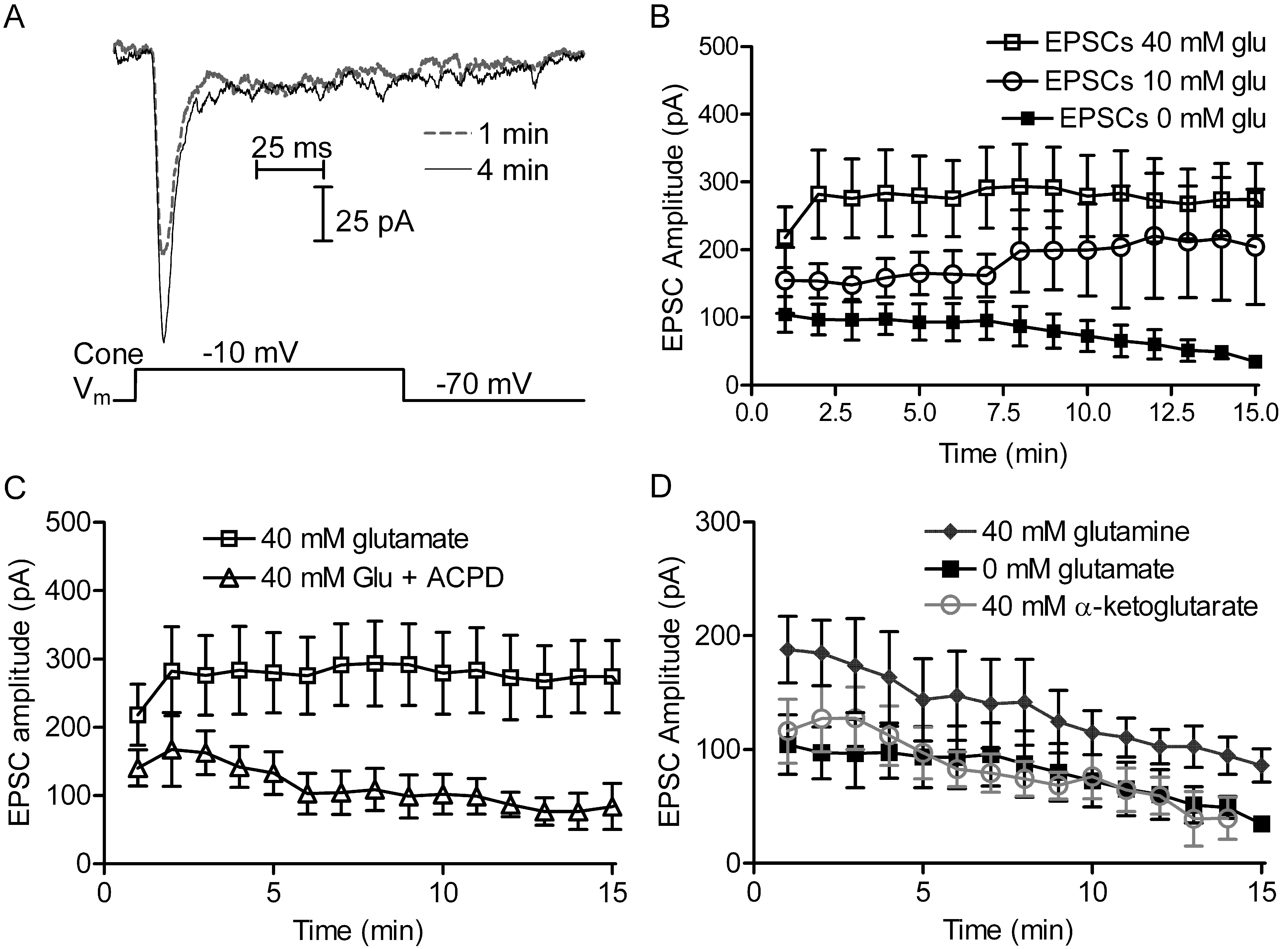
 Figure 1 of Bartoletti, Mol Vis 2011; 17:920-931.