Figure 5. The predicted secondary structures of the mutant and the wild type amino acid sequences. The predicted secondary structures
of the wild-type amino acid sequence (A) and the mutant amino acid sequence (B) is shown. The target sequences are labeled by the solid line, which indicate that the original α-helix is replaced by a
coil in the mutant type. Blue: helix; Yellow: sheet; Black: coil.
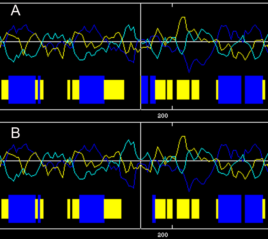
 Figure 5 of
Wang, Mol Vis 2011; 17:70-77.
Figure 5 of
Wang, Mol Vis 2011; 17:70-77.