Figure 6 of
Roshan, Mol Vis 2010; 16:887-896.
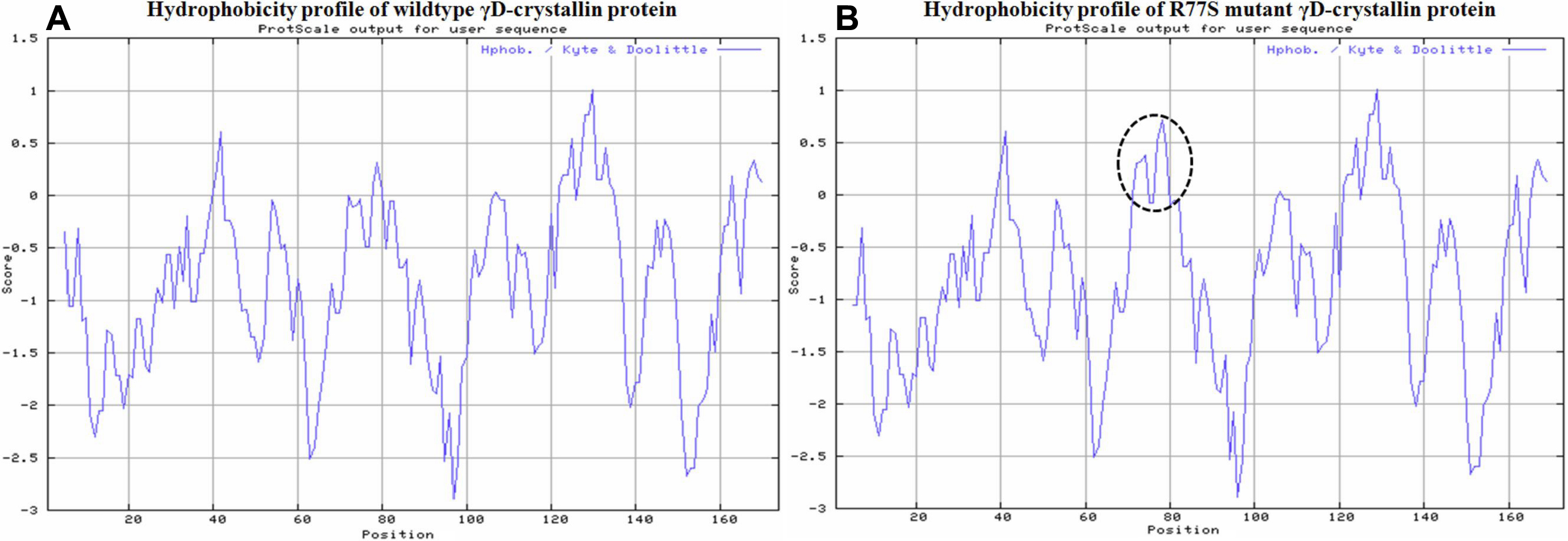
Figure 6.
Hydrophobicity profile of wild-type and R77S mutant γD-crystallin protein. Dotted circle represent the shift in the hydrophobicity around the mutant site. The prediction was done by ProtScale program at Expasy server.
 Figure 6 of Roshan, Mol Vis 2010; 16:887-896.