Figure 1. Photomicrograph showing a
septate, branching hypha (arrow) of Aspergillus flavus in
corneal scrape material from a patient with keratitis (Gram stain;
1,000×). Direct microscopic examination of corneal material by the
method of Gram staining revealed the presence of fungal hyphae.
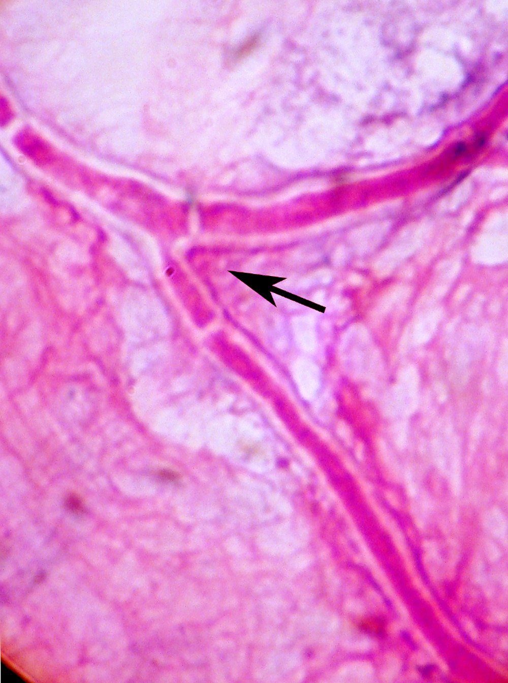
 Figure 1 of Leema, Mol Vis 2010; 16:843-854.