Figure 2. The retinoblastoma Y79 cell line
was treated with 5 µM Azacytidine (AZC) and the epithelial cell
adhesion molecule (Ep-CAM) expression was studied by flow cytometry
analysis (FC), Immunohistochemistry (IHC) and real time quantitative
reverse transcriptase polymerase chain reaction (Q-RT–PCR). A-E:
M1
represents the EpCAM percentage positivity in the FC graphs (solid
line). FC analysis demonstrated 47.73%, 44.2%, 42.96%, and 18.27% of
Y79 cells positive for Ep-CAM on days 5, 8, 10, and 15 of AZC
treatment, respectively. F-I: Immunohistchemistry pictures
stained with Di-amino benzidine (DAB) and was counter stained with
hematoxylin. All the pictures were captured at 20 times magnification.
EpCAM was not stained in Y79 cells not treated with AZC (F);
Strong membrane positive expression of Ep-CAM was seen in Y79 cells
treated AZC on 5th day (G); Strong membrane positivity of Ep-CAM
in the retinoblastoma primary tumor represented as positive control (H);
Y79
cells were stained negative when isotypic antibodies were used as
the negative control (I). Q-RT–PCR shows the mRNA expression of
Ep-CAM in Y79 cells after 5 days of AZC treatment (J). After day
5, Y79 cells were withdrawn from AZC exposure and subsequently passaged
in the complete medium up to 30 days, and Ep-CAM mRNA expression was
retained up to 21 days as follows: 210-fold, 132-fold, 56-fold, and
20-fold on days 8, 10, 15, and 20 days, respectively. The error bars
represents the standard deviation of triplicate values (J).
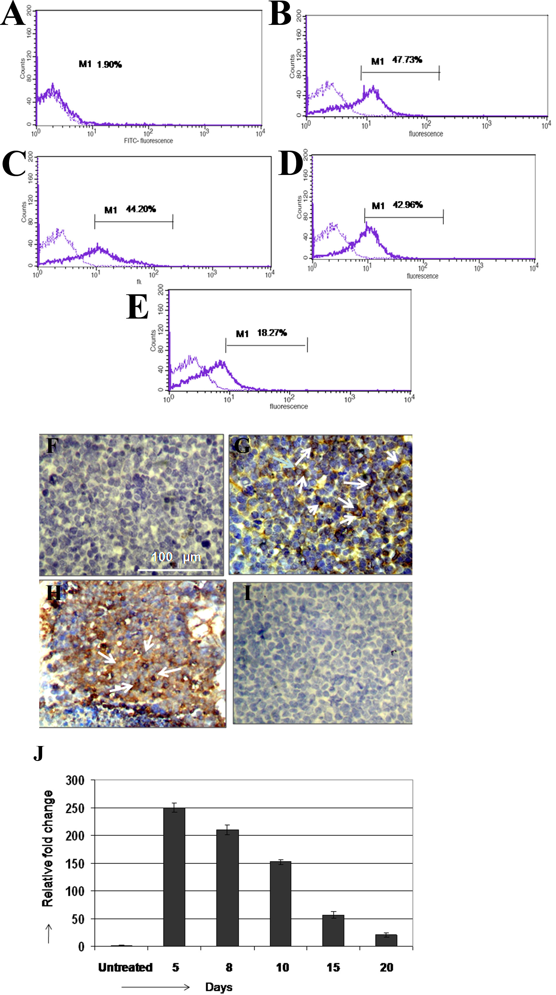
 Figure 2 of Mitra, Mol Vis 2010; 16:828-842.