Figure 10. Lysosomal morphology is changed after siramesine treatment. Note the large and heavily stained lysosomal structures in the
treated cells. Human lens epithelial cells (HLEC) was exposed to 30 µM of siramesine (B) or solvent (A) for 3 h at 37 °C before addition of the fluorogenic cathepsin B substrate Magic Red (RR-cresyl violet; red). Cell nuclei
are stained with Hoechst (blue). Confocal microscopy was performed 30 min after addition of the substrate. More than three
independent experiments were performed with similar results.
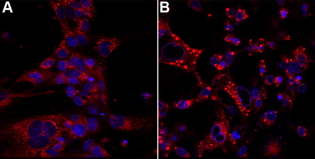
 Figure 10 of
Jonhede, Mol Vis 2010; 16:819-827.
Figure 10 of
Jonhede, Mol Vis 2010; 16:819-827.