Figure 3. Summary of previously reported TFAP2A mutations. A schematic of the TFAP2A gene is shown in A along with the genomic location of previously reported mutations. B and C show two different isoforms of the protein encoded by TFAP2A (AP2) along with the location of previously reported mutations at the protein level. The mutation in the current study is
indicated by red asterisk. PG: proline/glutamine rich domain, B: basic DNA binding domain, HSH: helix span helix domain.
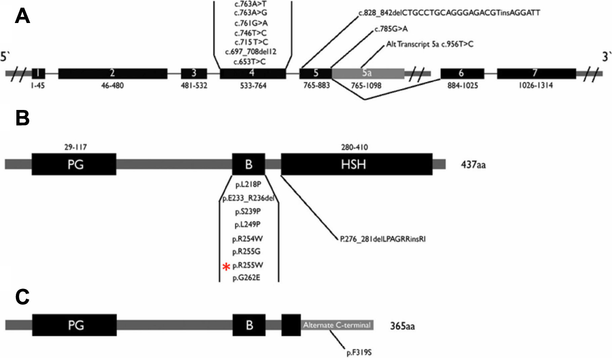
 Figure 3 of
Al-Dosari, Mol Vis 2010; 16:813-818.
Figure 3 of
Al-Dosari, Mol Vis 2010; 16:813-818.