Figure 1 of
Al-Dosari, Mol Vis 2010; 16:813-818.
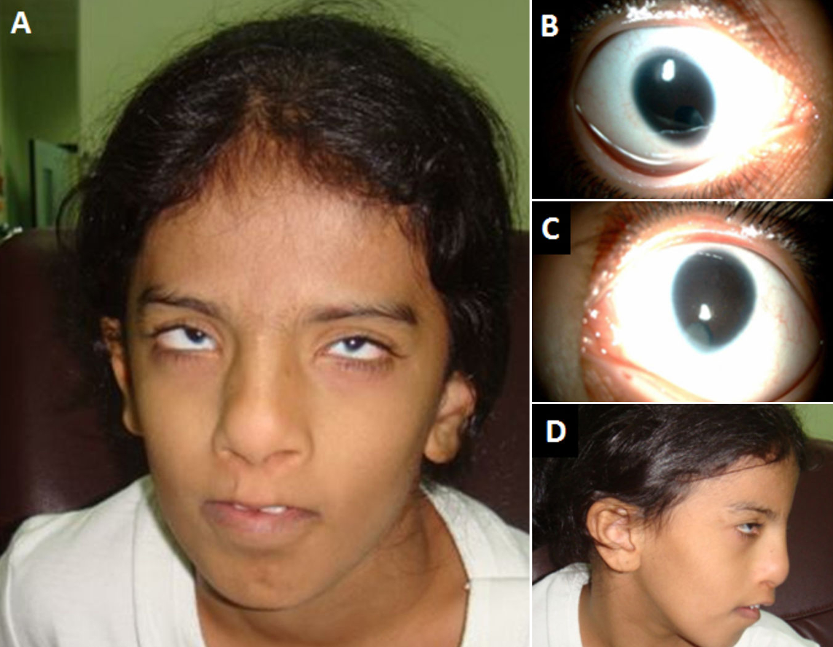
Figure 1.
Clinical photographs of the patient. Facial appearance with the typical pseudocleft of the upper lip is shown in
A
. Microcornea, inferonasal coloboma, and cataract are shown in
B
and
C
. Lateral facial profile is shown in
D
.
 Figure 1 of Al-Dosari, Mol Vis 2010; 16:813-818.