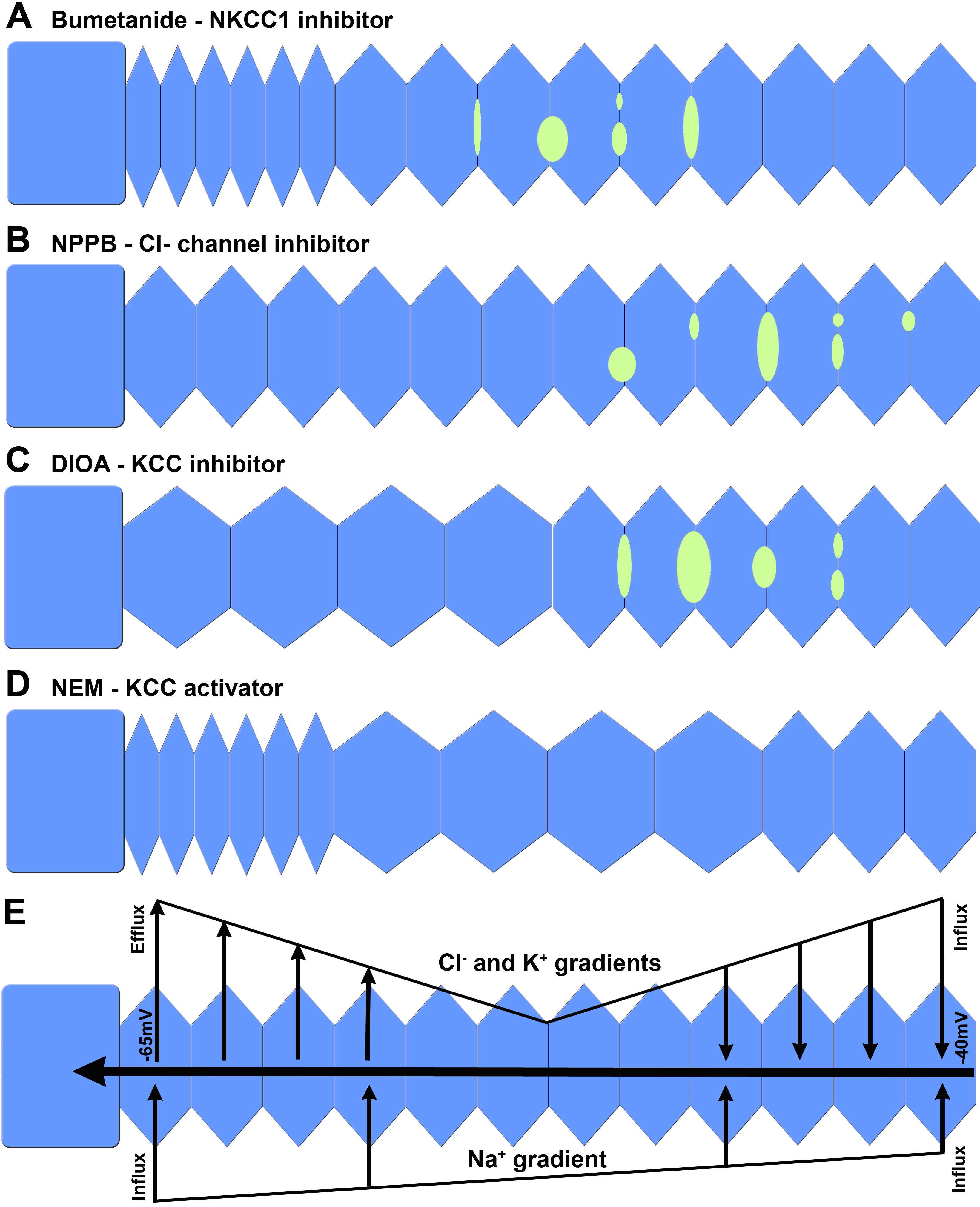
Figure 6. Modulation of ion transport
pathways has specific effects on fiber cell morphology in the rat lens
cortex. Schematic diagrams summarizing the distinctly different damage
phenotypes induced in the outer cortex of rat lenses organ cultured in
the presence of modulators of cation chloride cotransporters and Cl
−
channels for 18 h.
A: The NKCC inhibitor bumetanide (2 μM)
caused peripheral cell shrinkage and extracellular space dilations in
the deeper influx zone.
B: The Cl
- channel inhibitor
NPPB (10 μM) had no effect on peripheral fiber cells at this
concentration, but did induce extracellular space dilations between
deeper fiber cells.
C: The KCC inhibitor DIOA (10 μM) caused
peripheral cell swelling and deeper extracellular space dilations.
D:
The
KCC
activator
NEM (1 mM) caused shrinkage of peripheral fiber cells
but swelling of deeper fiber cells in the influx zone.
E: The
observed spatially distinct damage phenotypes can be explained by
differential changes in the electrochemical ion gradients for Cl
-
(E
Cl) and Na
+ (E
Na) that occur with
radial distance into the lens [
40] and their effects on the direction of ion
flux mediated by transporters in the two regions of the lens. In
peripheral fiber cells outwardly directed Cl
- gradients
favor Cl
- efflux mediated predominately by KCC [
11], while in deeper
fiber cells the lower transmembrane potential results in a shift in E
Cl
that favors influx mediated by KCC and Cl
- channels located
in this zone of the lens. In contrast E
Na is expected to
favor ion influx throughout the entire lens cortex.
 Figure 6 of Chee, Mol Vis 2010; 16:800-812.