Figure 5. Effects of NCC and NKCC
inhibitors on lens transparency and fiber cell morphology. Rat lenses
were organ cultured for 18 h in isotonic AAH in either the absence (A)
or
presence of the NCC inhibitor thiazide (B) or the NKCC
inhibitor bumetanide (C). Lens transparency was monitored by
dark field microscopy while fiber cell morphology was determined by
imaging equatorial sections labeled with the membrane marker WGA.
Culturing lenses in AAH only (A) or AAH + 10 μM thiazide (B)
had
no major effects on lens transparency (left panels) or fiber cell
morphology (right panels). C: Culturing lenses in AAH + 2 μM
bumetanide caused cortical opacification of the lens (left panel) that
was induced by damage to cortical fiber cell morphology (right panel). D-F:
High
powered images from the areas indicated (boxes) in A and C.
Fiber
cells in the lens periphery exhibited a marked shrinkage in the
presence of bumetanide (D) relative to that observed in the
absence of the inhibitor (E), while in the deeper influx zone
dilations of the extracellular space between fiber cells (F) was
observed in the presence of bumetanide.
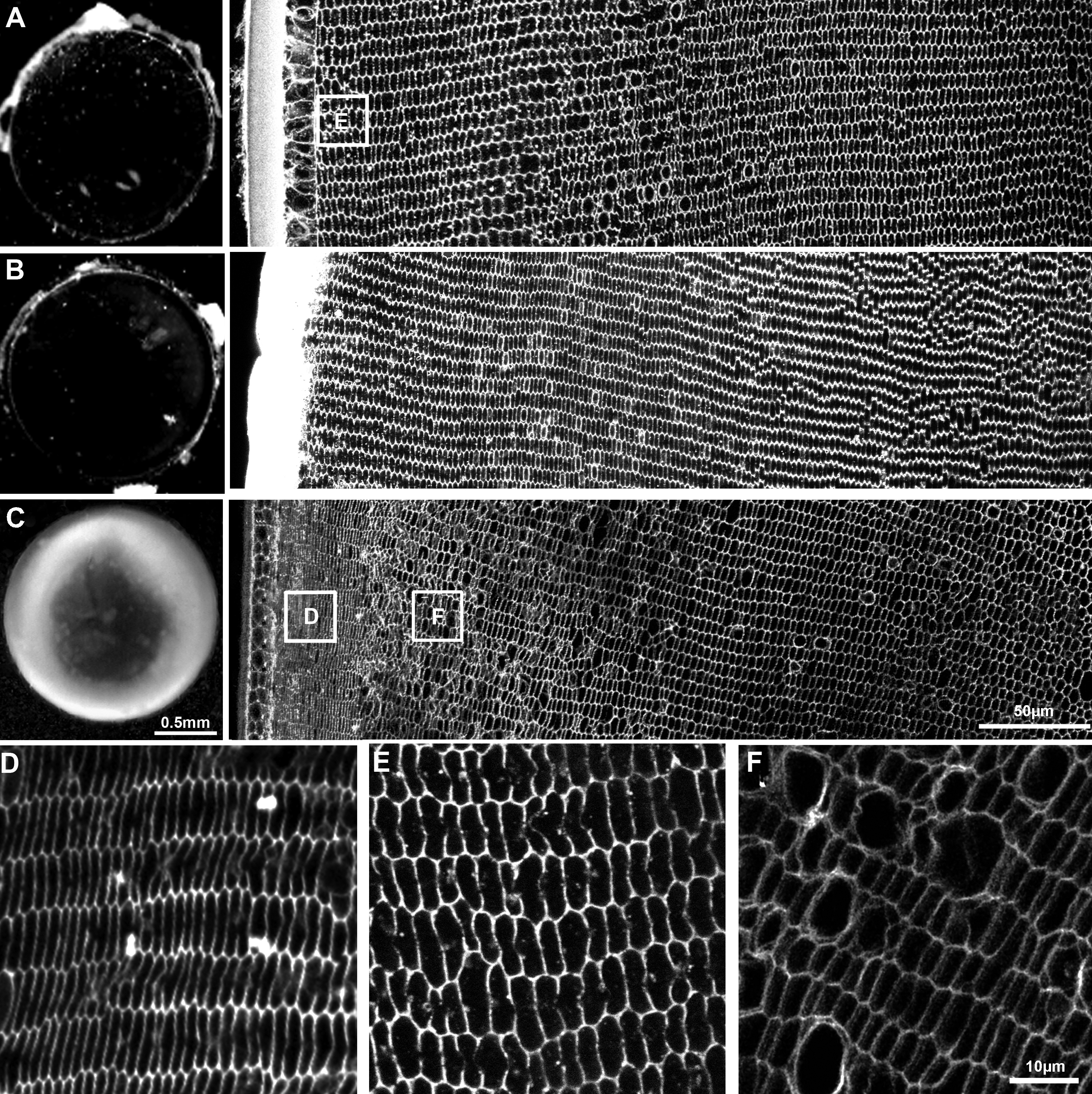
 Figure 5 of Chee, Mol Vis 2010; 16:800-812.