Figure 4. Example of scotopic
electroretinogram (ERG) in groups before bevacizumab (A),
anti-rat vascular endothelial growth factor (VEGF) antibody (B),
rat immunoglobulin G (IgG; C), and physiologic saline (D)
injection,
and 7 days after intravitreal injection of 5 µl of 3.75
mg/ml bevacizumab (E), 15 µg/ml anti-rat VEGF antibody (F),
1
mg/ml rat IgG (G), or physiologic saline (H). No significant
difference in b-wave amplitude was observed between bevacizumab,
anti-rat VEGF antibody, rat IgG, and physiologic saline injection
groups.
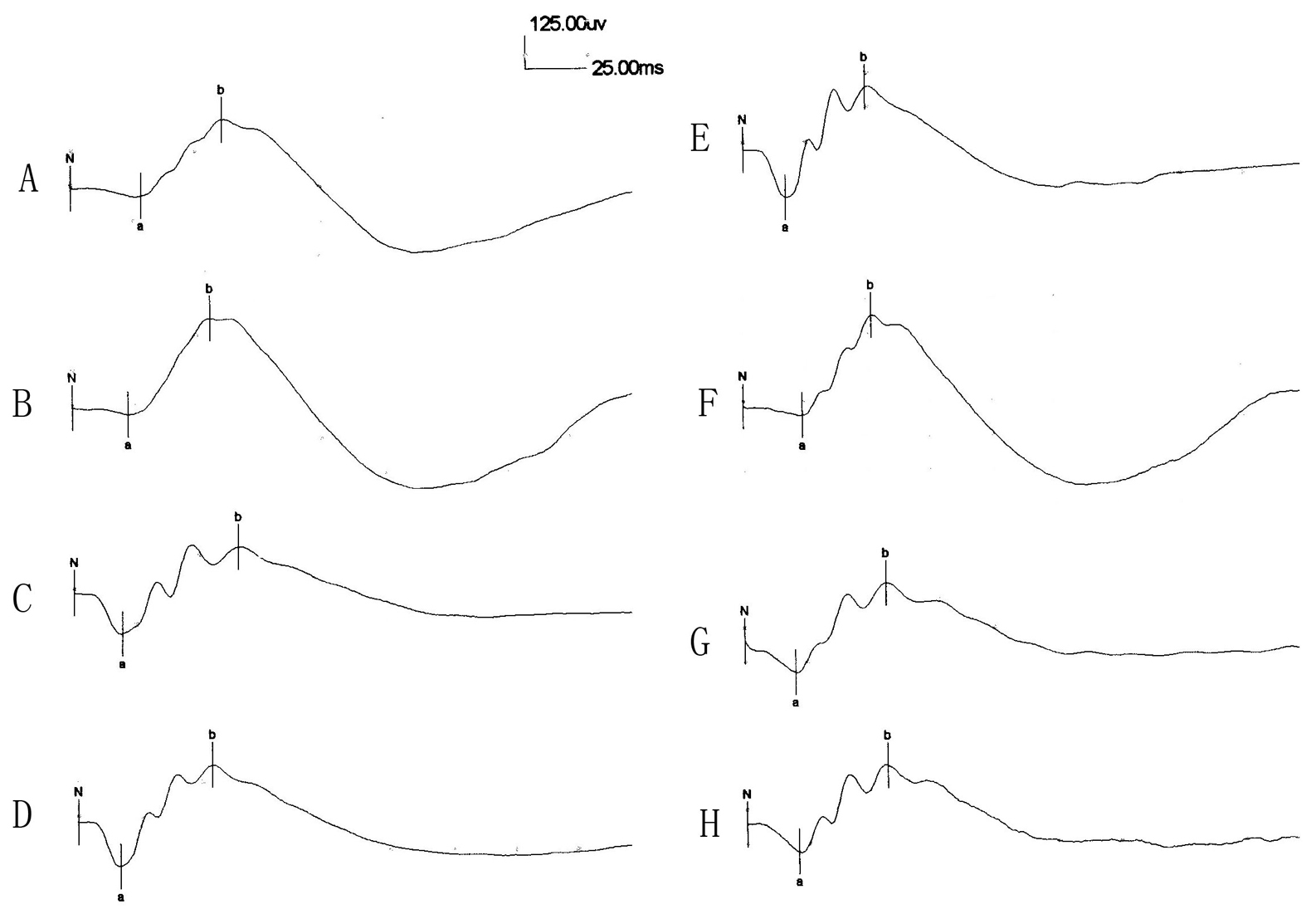
 Figure 4 of Guo, Mol Vis 2010; 16:793-799.