Figure 3. The histologic sections of the
retina with hematoxylin and eosin (HE) staining 7 days after
intravitreal injection of 5 µl of 3.75 mg/ml bevacizumab (A), 15
µg/ml anti-rat vascular endothelial growth factor (VEGF) antibody (B),
1
mg/ml rat immunoglobulin G (IgG; C), and physiologic saline (D).
E–H are the fluorescence images of glial fibrillary
acidic protein (GFAP)-immunolabeled sections 7 days after intravitreal
injection of bevacizumab (E), anti-rat VEGF antibody (F),
rat IgG (G), and physiologic saline (H). The scale bar
represents 20 µm.
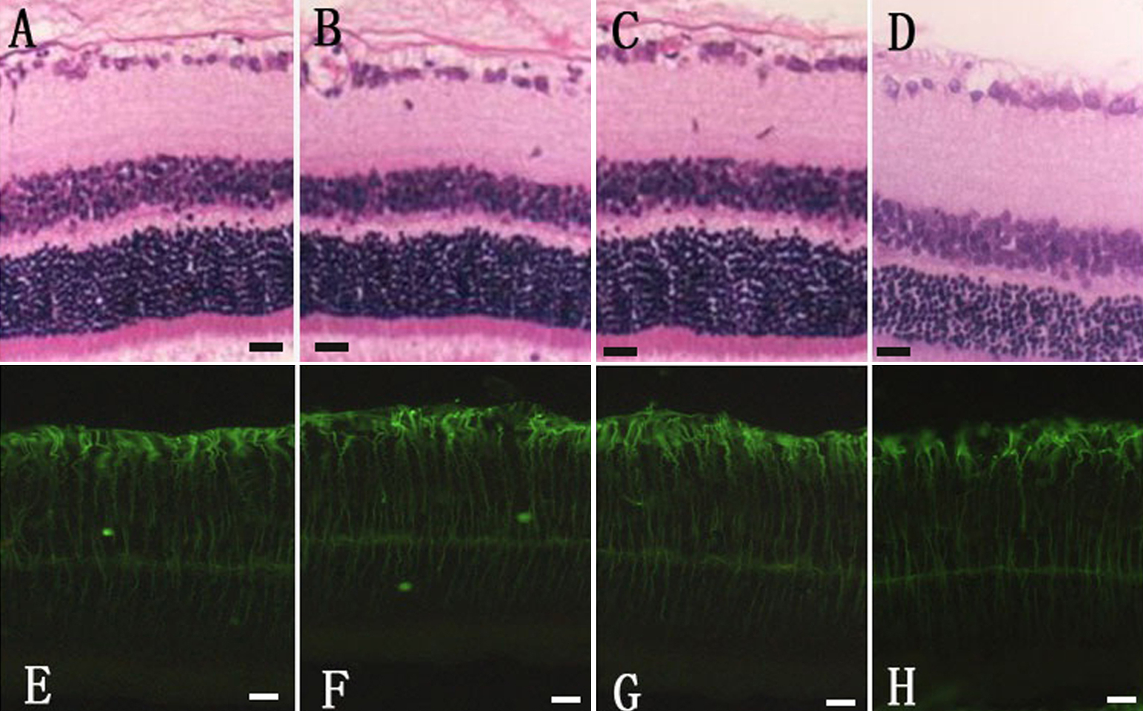
 Figure 3 of Guo, Mol Vis 2010; 16:793-799.