Figure 2. Methyl thiazolyl tetrazolium (MTT) assay showing optical densities of rat retinal Müller glial cells (RMGCs) after exposure
for 12, 24, 48, and 72 h to bevacizumab (0.1, 0.25, and 1 mg/ml), anti-rat vascular endothelial growth factor (VEGF) antibody
(0.1, 0.5 and 1 μg/ml), 1 mg/ml rat immunoglobulin G (IgG), and fresh medium as a control. Data were shown as the mean±SD
(n=5). The optical densities of RMGCs exposed for 48 and 72 h to anti-rat VEGF antibody at the concentrations of 0.5 μg/ml
and 1 μg/ml decreased remarkably. The optical densities of RMGCs were not affected after exposition to bevacizumab, IgG, and
fresh control medium. Each value represents the mean of three replicates. *, p<0.01 and +, p<0.05 differs from the control.
#, p<0.01 represents significant difference between various exposure time in 0.5 or 1 µg/ml ARVA group.
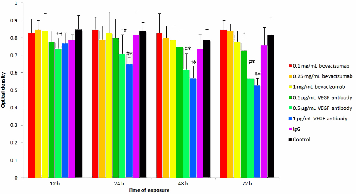
 Figure 2 of
Guo, Mol Vis 2010; 16:793-799.
Figure 2 of
Guo, Mol Vis 2010; 16:793-799.