Figure 1. Trypan blue exclusion assay showing the percentages of trypan blue-positive rat retinal Müller glial cells (RMGCs) after exposure
to bevacizumab (0.1, 0.25, and 1 mg/ml), anti-rat vascular endothelial growth factor (VEGF) antibody (0.1, 0.5 and 1 μg/ml),
1 mg/ml of rat immunoglobulin G (IgG), and control of fresh medium for 12, 24, 48, and 72 h . Data were shown as mean±SD (n=5).
The percentage of stained RMGCs at 48 and 72 h exposures to anti-rat VEGF antibody at the concentrations of 0.5 and 1 μg/ml
increased remarkably. The percentage of stained RMGCs was not affected after exposure to bevacizumab, IgG, and fresh control
medium. The experiment was performed at least three times with similar results. *, p<0.01 significantly differs from control.
#, p<0.01 represents significant difference between 48 and 72 h in 0.5 or 1 µg/ml ARVA group.
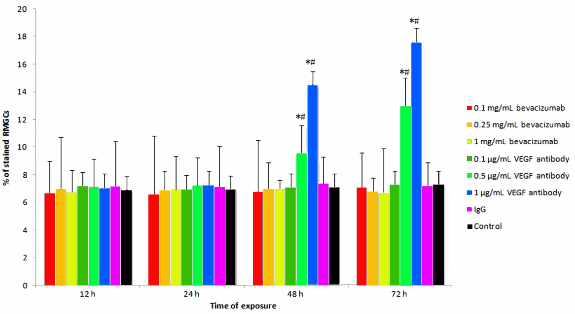
 Figure 1 of
Guo, Mol Vis 2010; 16:793-799.
Figure 1 of
Guo, Mol Vis 2010; 16:793-799.