Figure 5. Histopathological findings. A:
Hematoxylin and eosin staining at 20× reveals atrophied overlying
epithelium and Bowman’s membrane. The subepithelium and superficial
stroma are seen containing eosinophilic material. B: Amyloid
deposition was confirmed in the anterior and posterior stroma as
birefringence was seen on viewing under a polarized filter.
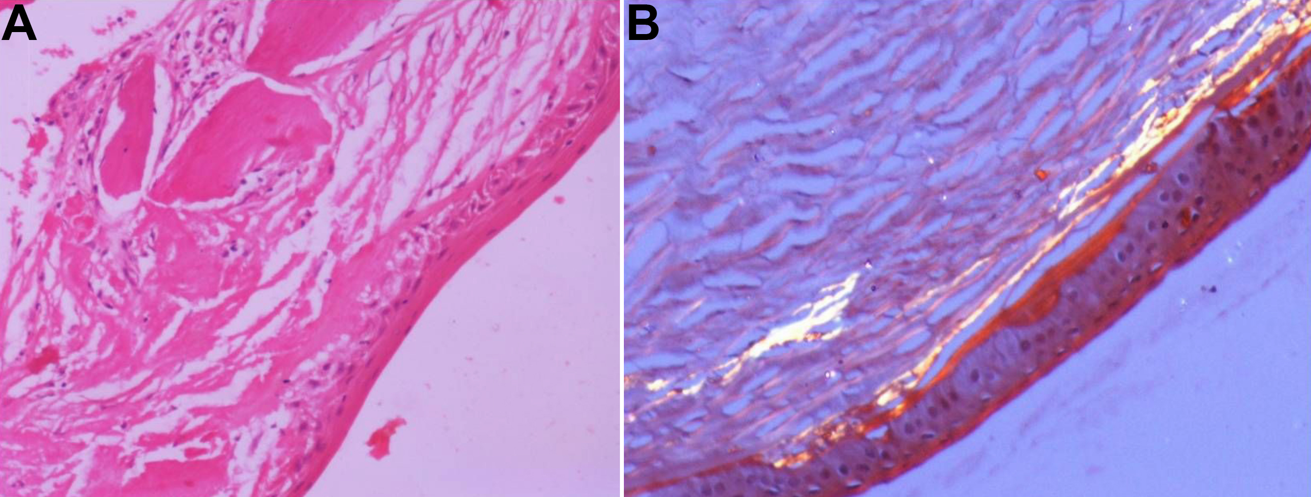
 Figure 5 of Paliwal, Mol Vis 2010; 16:729-739.