Figure 4 of
Paliwal, Mol Vis 2010; 16:729-739.
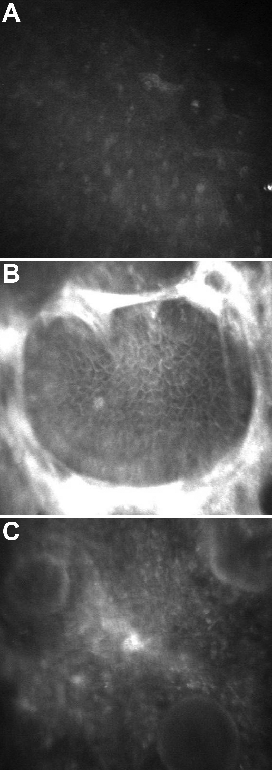
Figure 4.
In vivo confocal microscopic findings of the affected sibling. Normal superficial epithelium is shown (
A
). Basal epithelim with circumscribed hyperreflectivity is seen (
B
). Anterior stroma shows globular drop like structures (
C
).
 Figure 4 of
Paliwal, Mol Vis 2010; 16:729-739.
Figure 4 of
Paliwal, Mol Vis 2010; 16:729-739.