Figure 3. In vivo confocal microscopic findings of the affected individual. A: Superficial epithelial cells and B: basal epithelial cells with a well-demarcated cell border and the presence of reflective material are seen. C: Stroma/endothelium is not apparent.
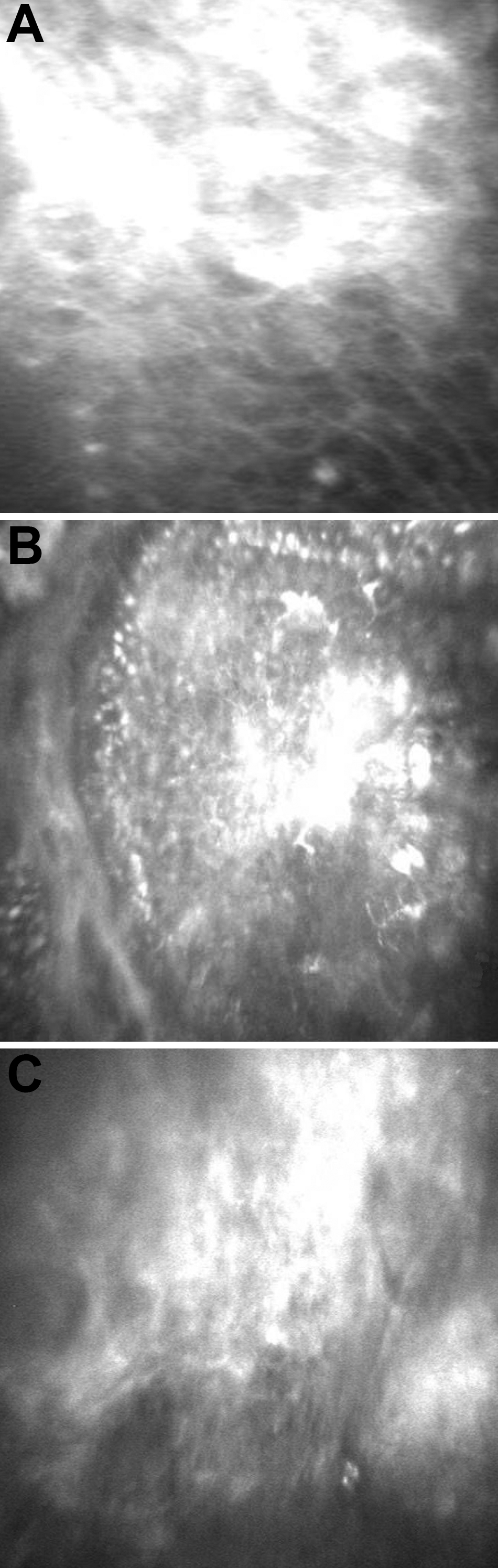
 Figure 3 of
Paliwal, Mol Vis 2010; 16:729-739.
Figure 3 of
Paliwal, Mol Vis 2010; 16:729-739.