Figure 2. Clinical photomicrographs of the
affected individuals of the family. The right eye (A) and left
eye (B) show advanced paracentral mulberry like deposition with
nodular opacification and neovascularization in the affected brother.
Right eye (C) of the affected sibling shows typical white
sub-epithelial nodules with mulberry pattern in the central cornea and
the left eye (D) of the same affected sibling shows band like
opacity in the interpalpebral area with neovascularization.
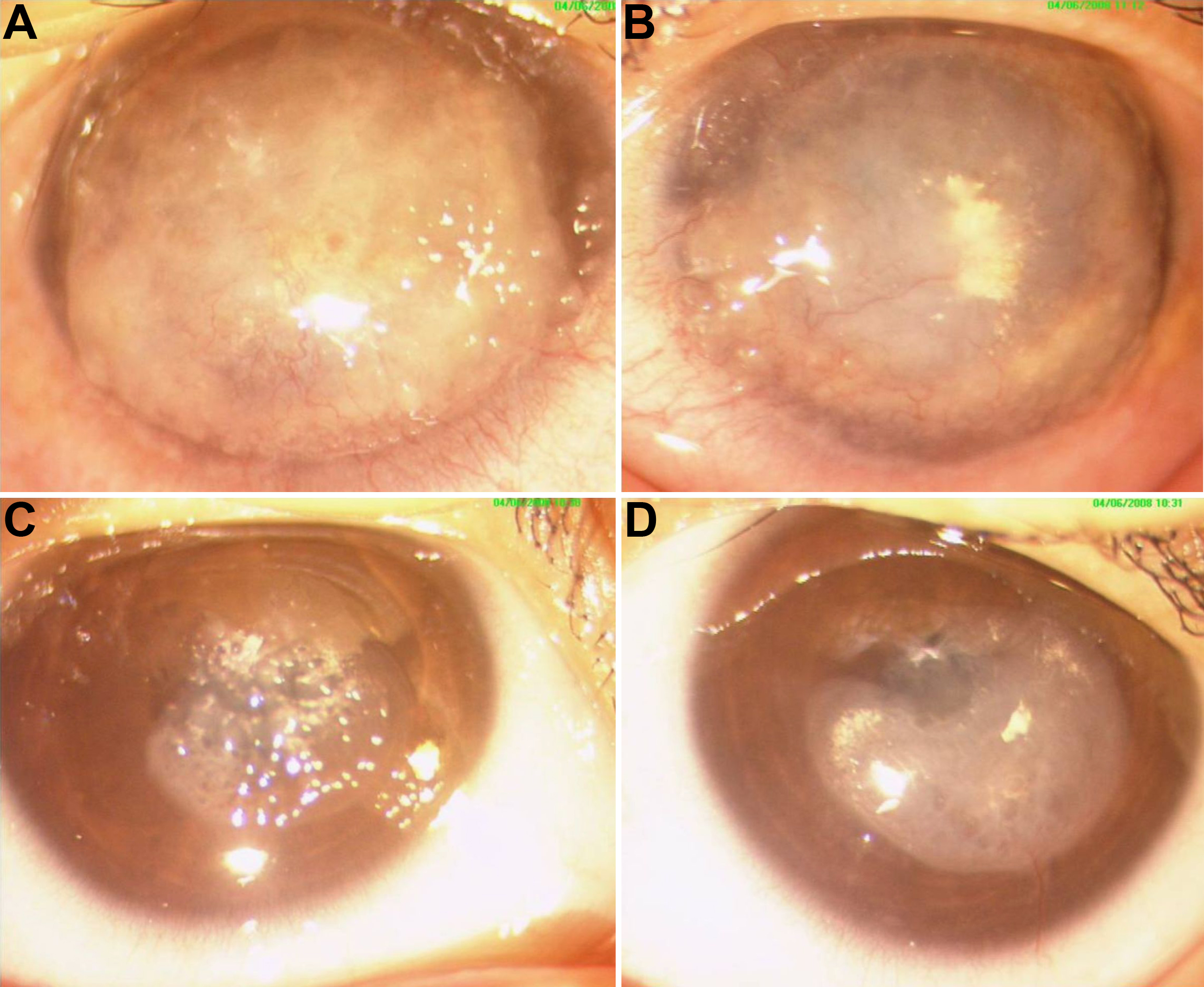
 Figure 2 of Paliwal, Mol Vis 2010; 16:729-739.