Figure 6. Representative
immunohistochemistry image showing collagen type IV staining of blood
vessels (red staining) in tissue sections of rabbit corneas collected
14 days after sterile water (A) or enalapril treatment (B).
The
images show angiogenesis in the central cornea region.
Enalapril-treated corneas showed fewer blood vessels. Nuclei are
stained blue with DAPI. The scale bar denotes 100 µm.
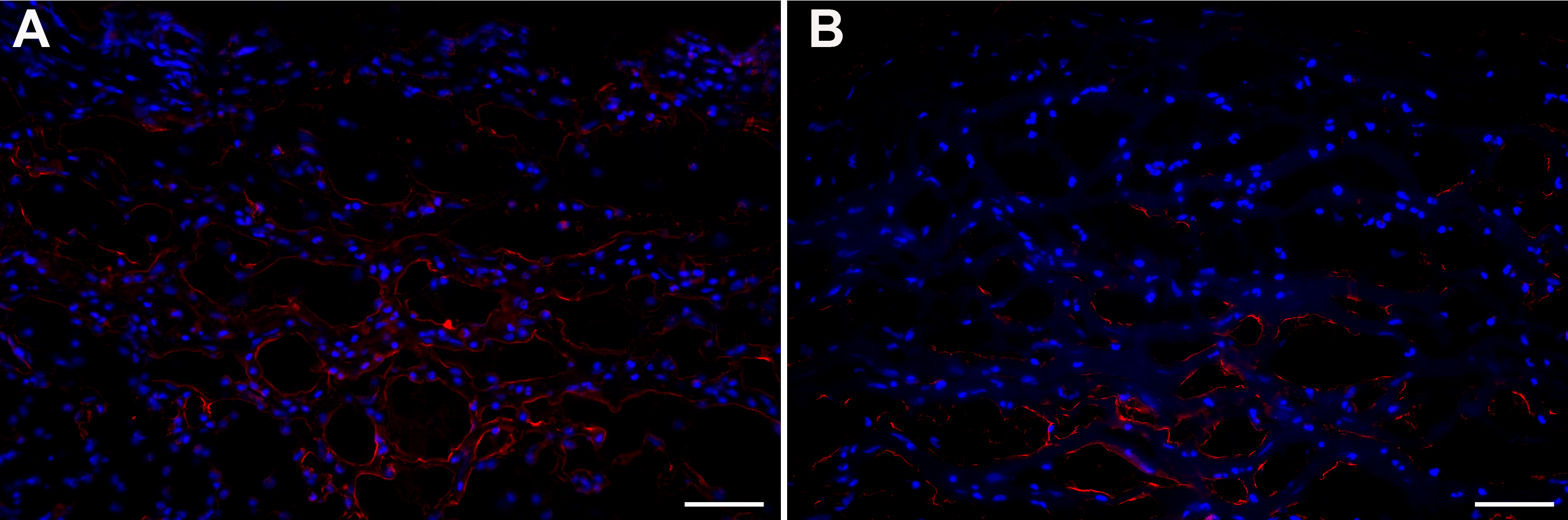
 Figure 6 of Sharma, Mol Vis 2010; 16:720-728.