Figure 3. Representative broad-beam (A)
and narrow-beam (B) slit-lamp biomicroscopy images of
VEGF-implanted rabbit corneas. Corneas did not show any cellular
infiltrate or edema in the area inferior to VEGF pellet (as shown by
arrow). A mild level of edema is noticeable around the area of VEGF
pellet. The scale bar denotes 2 mm.
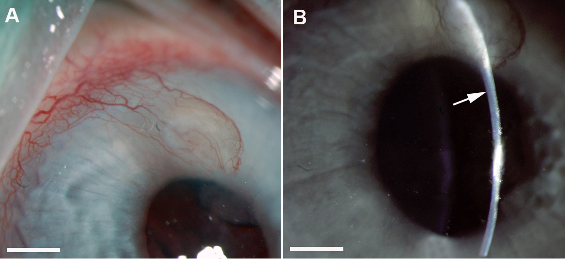
 Figure 3 of Sharma, Mol Vis 2010; 16:720-728.