Figure 5. The cytoskeleton architecture of glaucomatous trabecular meshwork cells is tangled. The organization of the filamentous actin
(F-actin; red fluorescence), and the distribution of vinculin (green fluorescence), the actin focal adhesion point on plasma
membrane, were observed using confocal microscopy. The glaucomatous trabecular meshwork cells have a more irregular actin
architecture and vinculin distribution compared to normal trabucular meshwork cells.
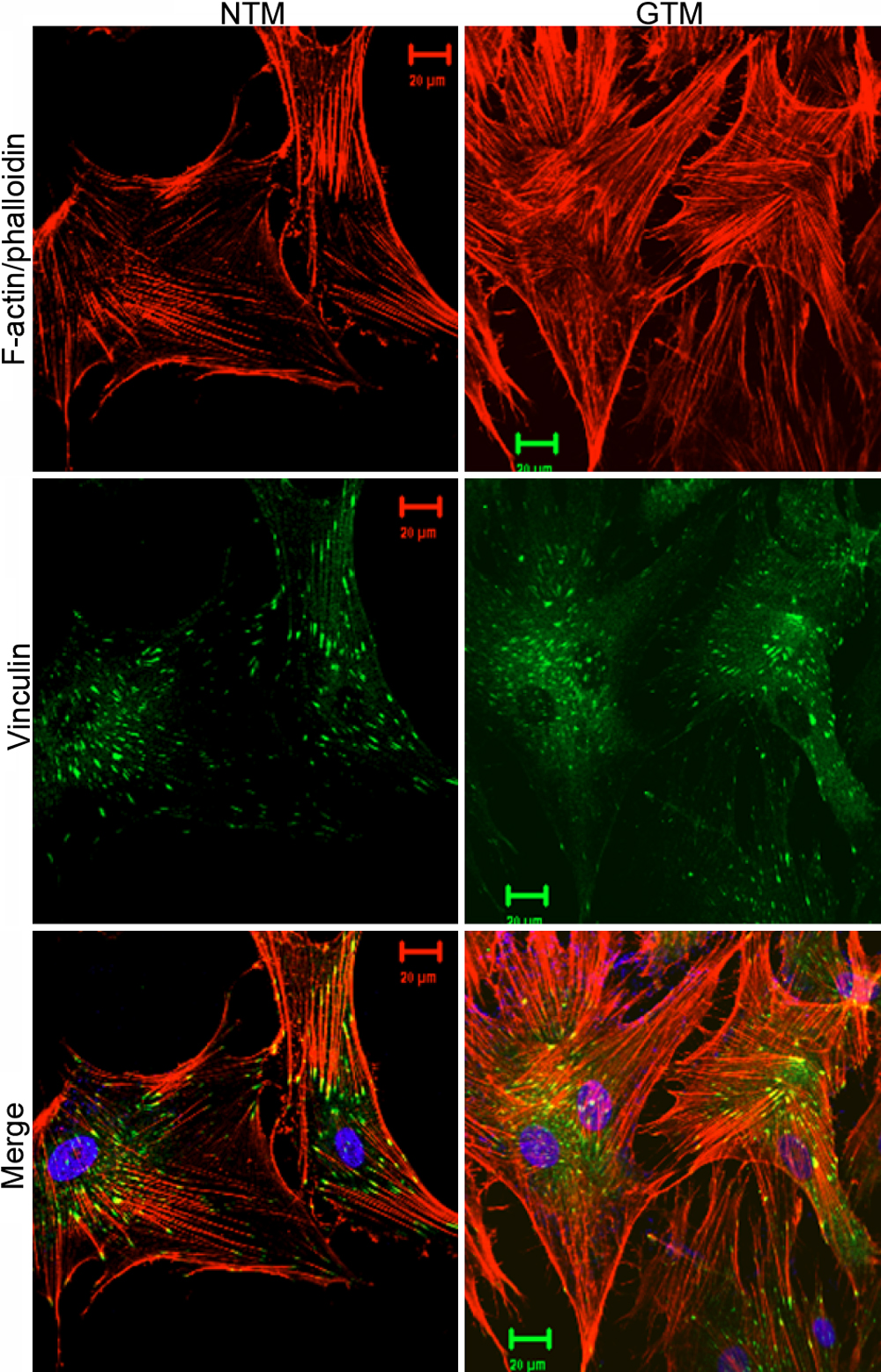
 Figure 5 of
Zhuo, Mol Vis 2010; 16:61-71.
Figure 5 of
Zhuo, Mol Vis 2010; 16:61-71.