Figure 6. Expression of fibulin-1 in human scleral fibroblasts (HSFs) visualized by indirect immunofluorescence. The nuclei were stained
with Propidium iodide dye (red: A, D, G), Dylight 488-conjugated secondary antibody was used for labeling with primary antibody (green: B, E, H). A–C: HSF cells were incubated in PBS without primary antibody as a negative control. D–F: HSFs were incubated in control medium. Fibulin-1 is weakly expressed in the cytoplasm. G–I: HSFs were incubated with retinoic acid (10−7 M) for 48 h. The expression of fibulin-1 in the cytoplasm of HSFs increased, and the morphology of HSFs changed. The original
magnification was 1,000× and the scale bar=10 μm.
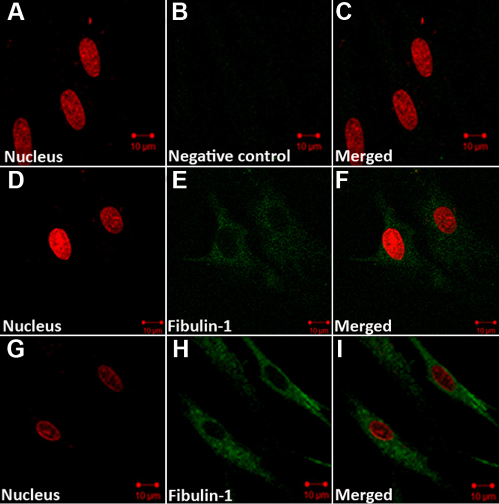
 Figure 6 of
Li, Mol Vis 2010; 16:689-697.
Figure 6 of
Li, Mol Vis 2010; 16:689-697.