Figure 4. Effect of all-trans-retinoic acid (RA) for both 24h and 48h on the expression of fibulin-1 protein levels in human scleral
fibroblasts (HSFs) using western blots. A: shows a western blot for fibulin-1 and GAPDH. No other bands were detected in the blot. Fibulin-1 was weakly expressed in
control HSFs and significantly enhanced when co-incubated with 10−7 M of RA. B: Bar graphs show changes in protein expression (mean±SD, n=3) where density values were compared to GAPDH density. The asterisk
indicates a significant difference relative to the control (p<0.05).
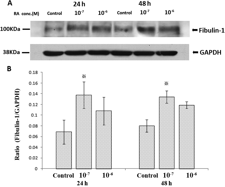
 Figure 4 of
Li, Mol Vis 2010; 16:689-697.
Figure 4 of
Li, Mol Vis 2010; 16:689-697.