Figure 1. Expression and localization of fibulin-1 and aggrecan in guinea pig sclera. Pigmented guinea pigs were fed all-trans-retinoic
acid (RA) or peanut oil (PO) for 5 days, then scleral sections were double labeled with antibodies for fibulin-1 (green) and
aggrecan (red). Hoechst 33342 dyed the nucleus (blue: A, E, J). A–D: PBS was used instead of primary antibody as a negative control. Fibulin-1 was localized in scleral extracellular matrices
in parallel with the distribution of aggrecan. E–H: Scleral tissue was taken from control animals fed vehicle (PO). Aggrecan was arranged between collagen fibrils and collagenous
lamellae and dispersed in the outer layer of sclera. Fibulin-1 staining was weaker and surrounded several scleral fibroblasts.
I–L: Scleral tissue was taken from animals fed RA. Aggrecan staining became weak, especially in the middle lamella, while fibulin-1
staining became substantially stronger. The original magnification was 400× and the scale bar=20 μm.
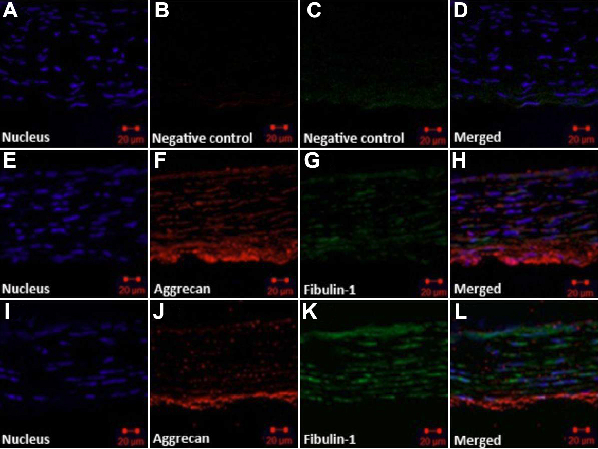
 Figure 1 of
Li, Mol Vis 2010; 16:689-697.
Figure 1 of
Li, Mol Vis 2010; 16:689-697.