Figure 5. Ocular manifestations in the
proband. The pictures show the aspects of the ocular anterior segment
as well as of the lens of the patient. The proband had microcornea,
microphthalmia, congenital corneal leukoma, iris dysplasia, and
anterior polar cataracts in both eyes. In A (right eye) and B
(left eye), arrowheads point to adherent corneal leucoma and arrows
point to anterior polar cataracts.
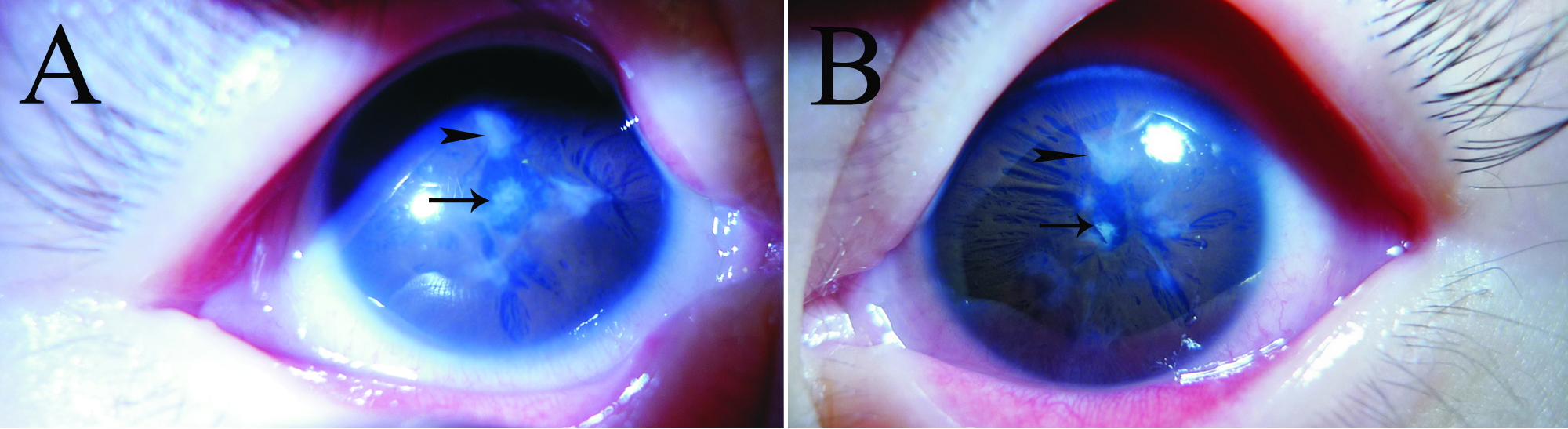
 Figure 5 of Jia, Mol Vis 2010; 16:676-681.