Figure 3. Immunolocalization of transient
receptor potential vanilloid channels, TRPV5 and TRPV6, in frozen
sections of native human retinal pigment epithelium/choroid tissue.
Tissue orientation in all panels shows the retinal pigment epithelium
(RPE) monolayer above the choroid tissue. In the fluorescent images
(left two panels), TRPV5 immunoreactivity (A) and TRPV6
immunoreactivity (B) appear green; nuclei (red) were visualized
with TO-PRO-3; corresponding differential interference contrast (DIC)
images (panels on the right) show the pigmented RPE monolayers. In A,
the
anti-TRPV5 antibody used was KL362 (affinity-purified polyclonal
antibody, final concentration 2.5 µg/ml). In B, the anti-TRPV6
antibody used was KL364 (affinity-purified polyclonal antibody, final
concentration 2.5 µg/ml). The secondary antibody used in both panels
was antirabbit immunoglobulin G (IgG) conjugated to Alexa 488 (final
dilution 1:500). Down arrows in both panels identify prominent staining
on or just below the apical plasma membrane; up arrows identify
prominent staining along the basal plasma membrane. Sections are from a
47-year-old Caucasian female donor. The pattern of staining in these
panels is representative of staining observed on sections from four
different donors (three female, one male, ranging in age from 47 to 79
years). Scale bars in all panels represent 10 µm.
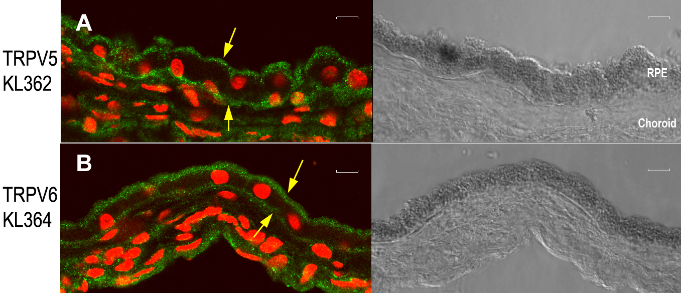
 Figure 3 of Kennedy, Mol Vis 2010; 16:665-675.