Figure 12. Sequence chromatogram of the transmembrane protein 126A gene mutation in the optic atrophy family, and their corresponding
normal sequence. A: This panel shows the chromatogram of a control sample with wild-type allele. B: This panel shows the chromatogram of the mother (III:1) with the heterozygous transmembrane protein 126A gene (TMEM126A) mutation (c.163C>T). C: This panel shows the chromatogram of an affected individual (IV:1) with the homozygous TMEM126A variant (c.163T). The black framed box indicates the mutation position.
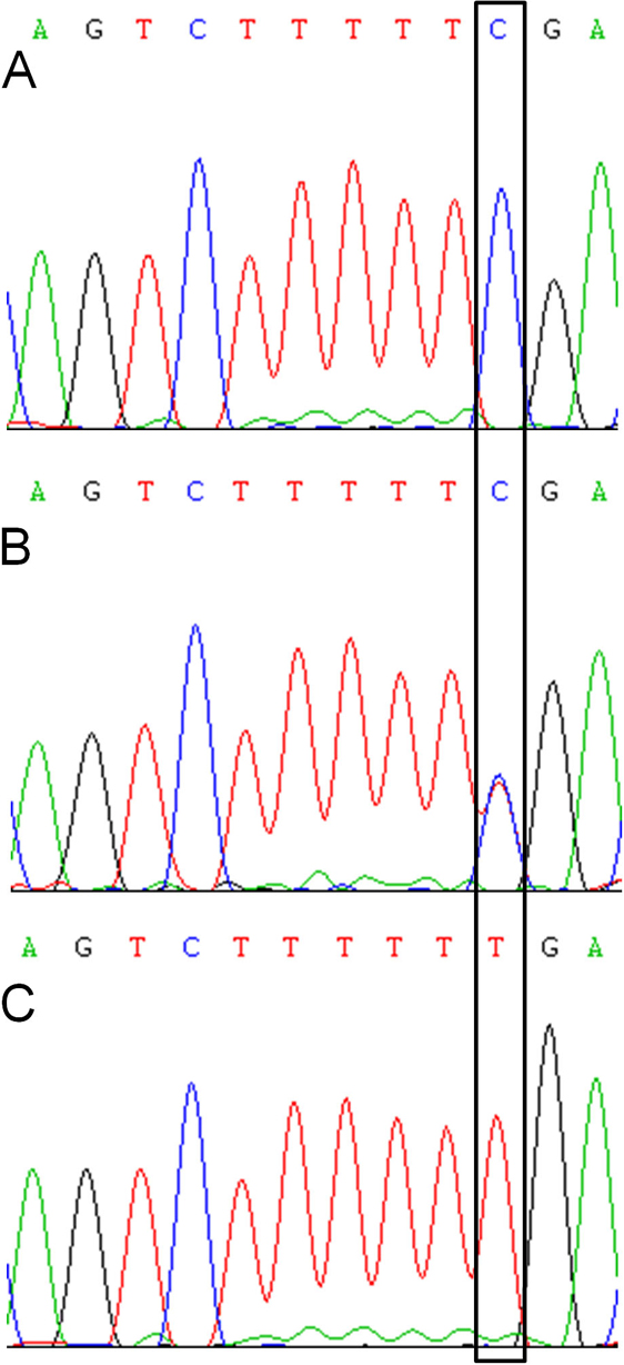
 Figure 12 of
Meyer, Mol Vis 2010; 16:650-664.
Figure 12 of
Meyer, Mol Vis 2010; 16:650-664.