Figure 2. Cell identification results by phase-contrast microscopy and RT–PCR. A: Confluent cultures of human corneal epithelial cells expressing K3 (keratin 3). B: Human corneal stromal fibroblast cells expressing Vim (vimentin). C: HCECs expressing Col VIII (collagen VIII) and NSE (neuron-specific enolase).
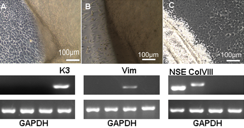
 Figure 2 of
Lu, Mol Vis 2010; 16:611-622.
Figure 2 of
Lu, Mol Vis 2010; 16:611-622.