Figure 5. Subretinal expression of
aquaporin 1 during experimental autoimmune uveitis. Retina was
submitted to immunofluorescent staining for aquaporin 1 (green; A).
Cell
nuclei were stained with DAPI (blue, B). C
corresponds to merge image. The arrow shows the cell nuclei of the RPE
cells monolayer. RPE represents retinal pigmented epithelium and CC
represents choriocapillaris. Magnification is 400×.
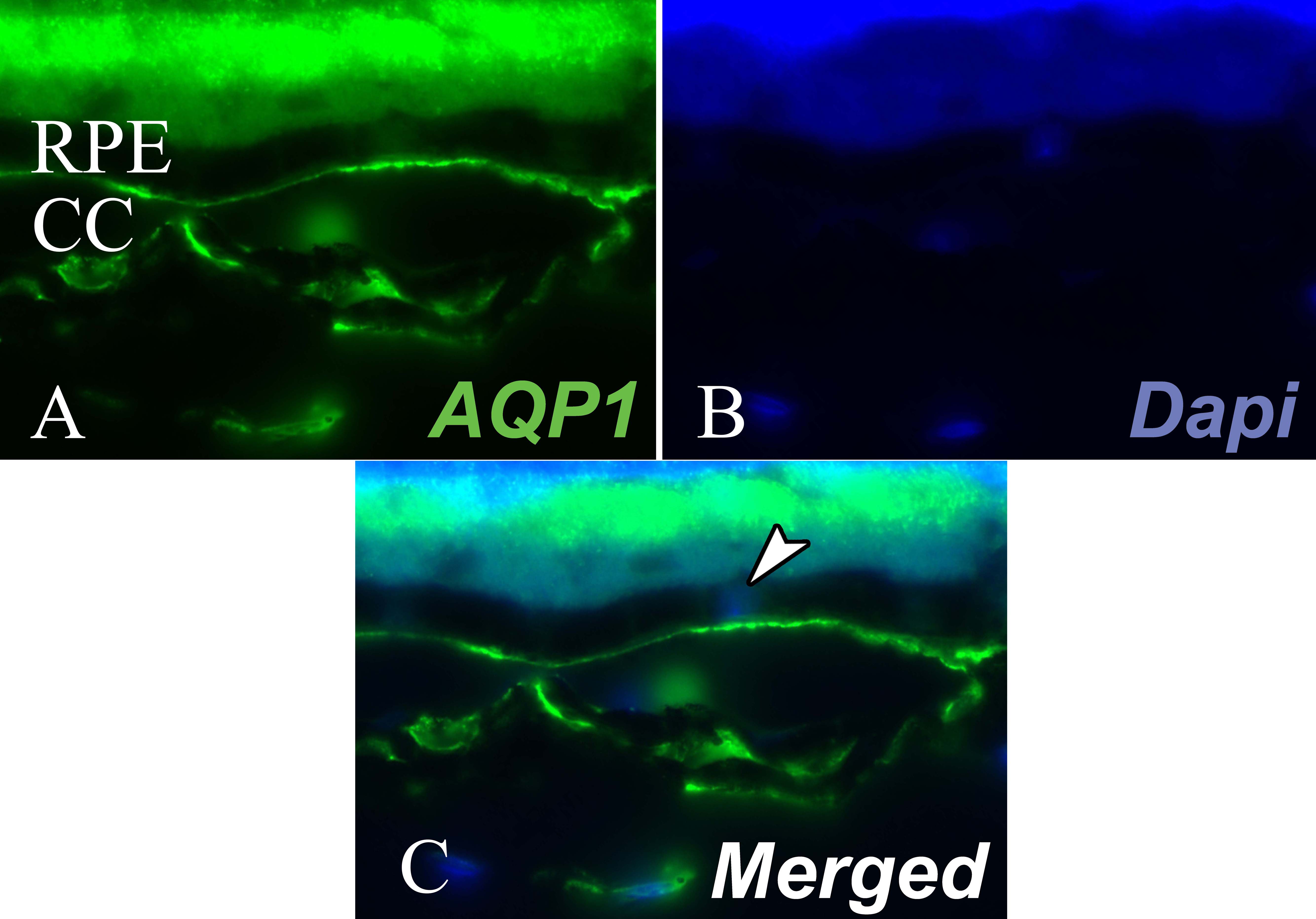
 Figure 5 of Motulsky, Mol Vis 2010; 16:602-610.