Figure 3. Retinal expression of aquaporin
1 and glial fibrillary acidic protein during experimental autoimmune
uveitis. The images illustrate retina without specific lesions (A, B,
and
C), with vasculitis (D, E, and F) or with
intraretinal inflammatory infiltrate (G, H, and I).
Retina was submitted to immunofluorescent staining for aquaporin 1
(AQP1; in green; A, D, and G), or to glial fibrillary
acidic protein (GFAP; in red; B, E, and H). Cell nuclei
were stained with DAPI (blue). C, F, and I
correspond to merged images. ILM represents inner limiting membrane;
OLM represents outer limiting membrane; and RPE represents retinal
pigmented epithelium. Magnification is 60×.
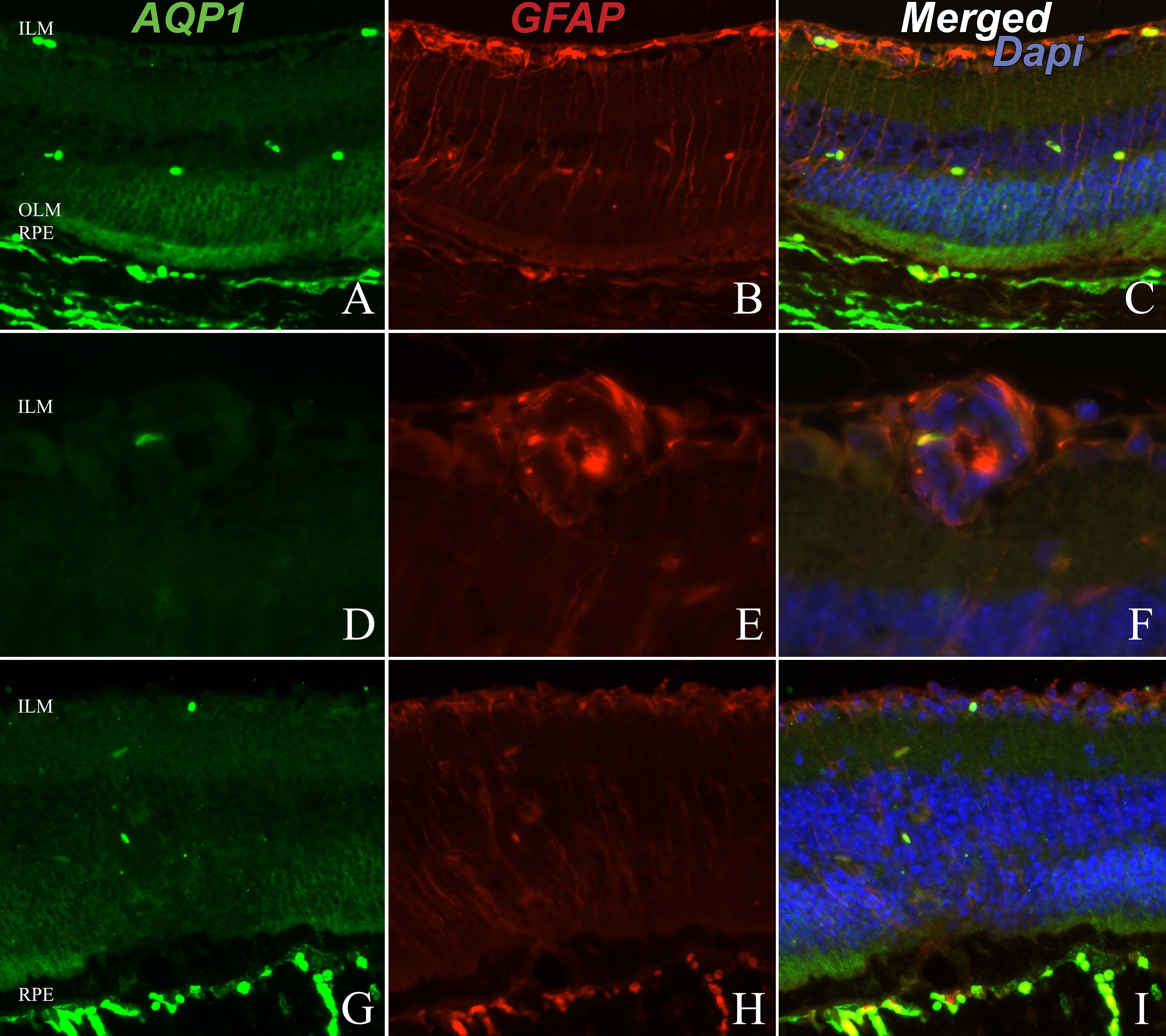
 Figure 3 of Motulsky, Mol Vis 2010; 16:602-610.