Figure 1. Expression of aquaporin 1,
aquaporin 4, and glial fibrillary acidic protein in normal mouse
retina. Normal mouse retina was submitted to hematoxylin and eosin
staining (A), or immunofluorescent staining for aquaporin 1
(AQP1; B), aquaporin 4 (AQP4; C), and glial fibrillary
acidic protein (GFAP; D). ILM represents inner limiting
membrane; GCL represents ganglion cell layer; IPL represents inner
plexiform layer; INL represents inner nuclear layer; OPL represents
outer plexiform layer; ONL represents outer nuclear layer; OLM
represents outer limiting membrane; IS represents inner segments of the
photoreceptors; OS represents outer segments of the photoreceptors; RPE
represents retinal pigmented epithelium. Magnification is 200×.
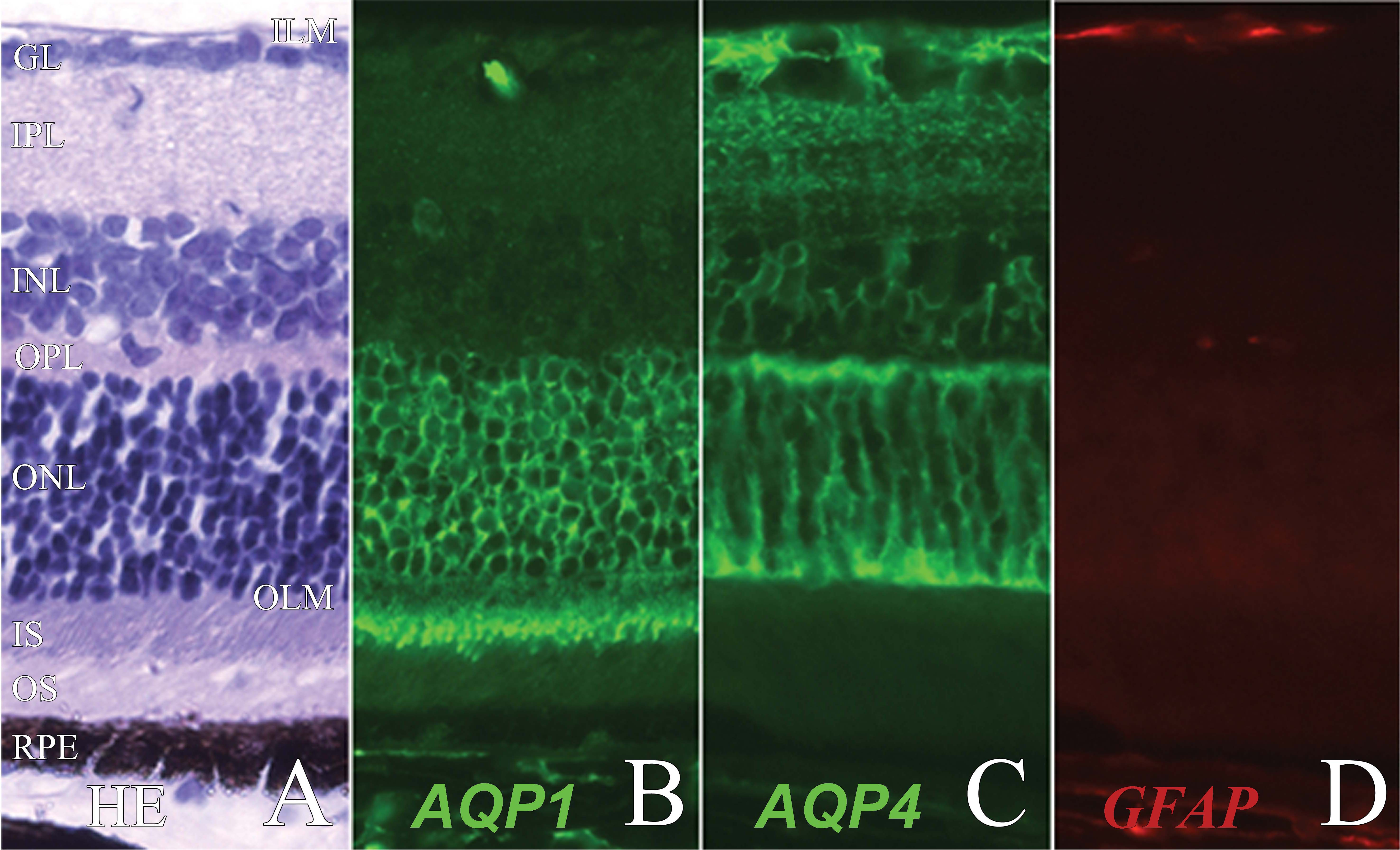
 Figure 1 of Motulsky, Mol Vis 2010; 16:602-610.