Figure 2. Effects of epigallocatechin
gallate (EGCG) on retinal pigment epithelium (RPE) cell migration. A,
B: Epigallocatechin gallate inhibits platelet-derived growth
factor (PDGF)-BB-induced RPE cell migration. Transwell inserts were
coated with fibronectin. Human RPE cells were seeded in the upper
chamber in the presence of vehicle or EGCG. The inserts were assembled
in the lower chamber, which was filled with serum-free ([−]PDGF-BB; A)
or
PDGF-BB-containing
medium ([+]PDGF-BB; B) and preincubated
with a vehicle or EGCG on the polycarbonate filter of the insert for
30 min. PBS, without PDGF-BB or EGCG, served as the control (A,
left).
Human
RPE cells that migrated to the underside of filter
membrane were photographed and counted in high-power field (HPF,
magnification, 100×) under a phase-contrast light microscope. The scale
bar represents 100 μm. The black spots are the pores of the
Transwell membrane and the grayish fusiform cells are the ARPE cells. C:
Quantitative
analysis
of migrated cells. Twenty HPFs were counted in
each migration. All experiments were conducted in duplicate and similar
results were obtained at least two to three times. Data are presented
as percent of control (the first unfilled bar, PBS only) in cell
counts. *p<0.05 significantly differs from PDGF-BB-stimulated cells
(the first filled bar).
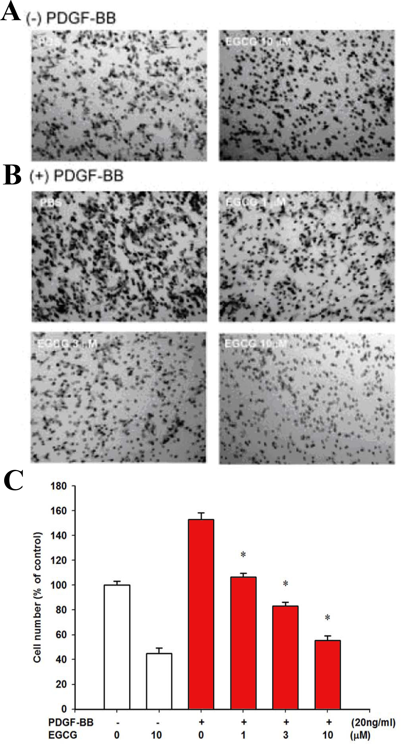
 Figure 2 of Chan, Mol Vis 2010; 16:586-595.