Figure 7. Three months after lesioning
both central and peripheral injuries caused by photocoagulation (PC)
were repaired with new retinal pigmented epithelium (RPE) cells that
expressed RPE specific markers. A: Otx2 expression marking RPE
cells was continuous across the lesion in whole mounted preparations.
The scale bar here represents 100 µm. B: Reveals that repaired
regions were hypopigmented with many RPE cells either lacking pigment
completely or containing reduced pigment levels. C: Otx2
expression in RPE cells was also continuous across lesions when they
were viewed in section. This image shows the location of these cells in
relation to the choroidal capillaries (CHO). The scale bar is 25 µm and
the image also contains a low power figure of the region. D:
The RPE tight junction protein ZO-1 (red) was also present across the
lesion site. The nuclei here are shown in blue. Unexpectedly,
photoreceptor outer segments (green) remained adhered to the repaired
tissue. Their location here demarcates the lesion site. The scale bar
here represents 50 µm. E: Shows a high power image of a lesion
site revealing Otx2 expression and the brighter cells inside due to the
absence of melanin. The scale bar here is 20 µm. F: A
transverse section through the repaired region of the RPE. The green
outer segments (OS) are indicated by the upper arrow. The lower arrow
head reveals the ZO-1 (red). The location of the CHO is also indicated
under the other tissues. Nuclei are blue. The scale bar is 20 µm.
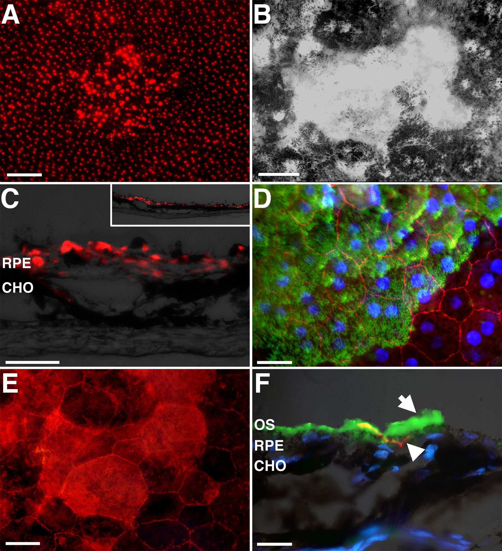
 Figure 7 of Lundh von Leithner, Mol Vis 2010; 16:570-581.