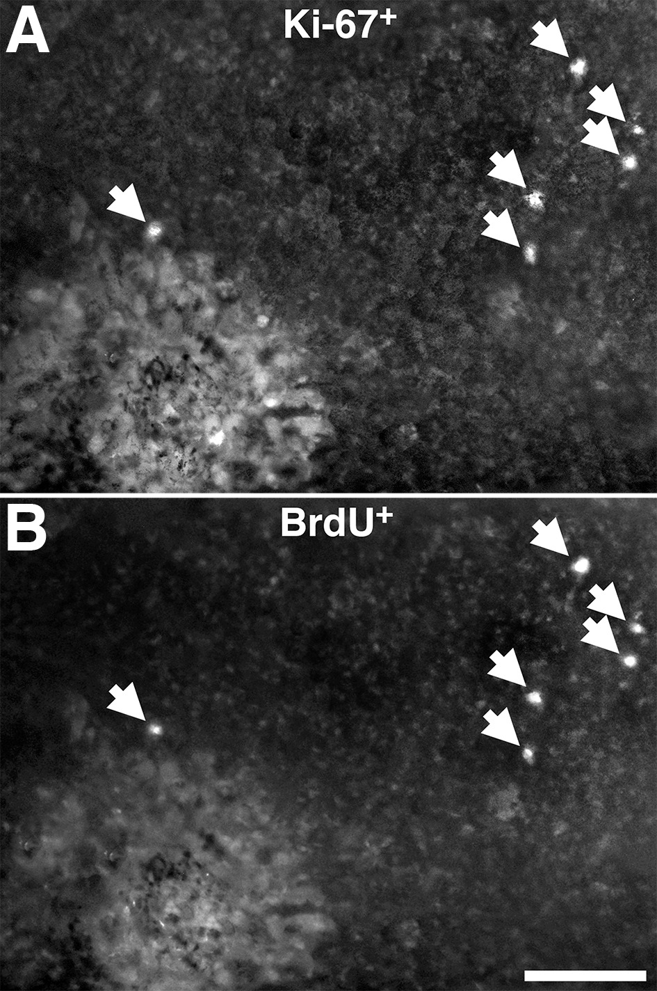
Figure 5. Double labeling combining Bromodeoxyuridine (BrdU) and Ki67 protocols 72 h following photocoagulation (PC) lesioning of the
RPE with one central and one peripheral lesion.
A shows labeling for Ki67 and
B for BrdU in the same tissue. Arrows indicate cell labeled with both markers. The data revealed extensive co-localization
of both labels, confirming that many of the cells that were positive for Ki67, and as such in the cell cycle, were also positive
for BrdU, confirming that full cell division had taken place. More cells positive for Ki67 and BrdU were always found in the
periphery than in the center, reflecting patterns found in
Figure 3 and
Figure 4 (data not shown). The scale bars represent 100 µm.
 Figure 5 of
Lundh von Leithner, Mol Vis 2010; 16:570-581.
Figure 5 of
Lundh von Leithner, Mol Vis 2010; 16:570-581.