Figure 2. Immunostaining of retinal pigment epithelium lesion site 72 h after injury. A: Immunoreactivity for activated caspase-3 in lesioned retinal pigment epithelium cells demonstrated that at this stage cell
death was still a feature of the tissue. B: Expression of metalloproteinase marker 2 was consistent with the process of extracellular degradation and tissue remodeling.
Scale bar represents 50 µm.
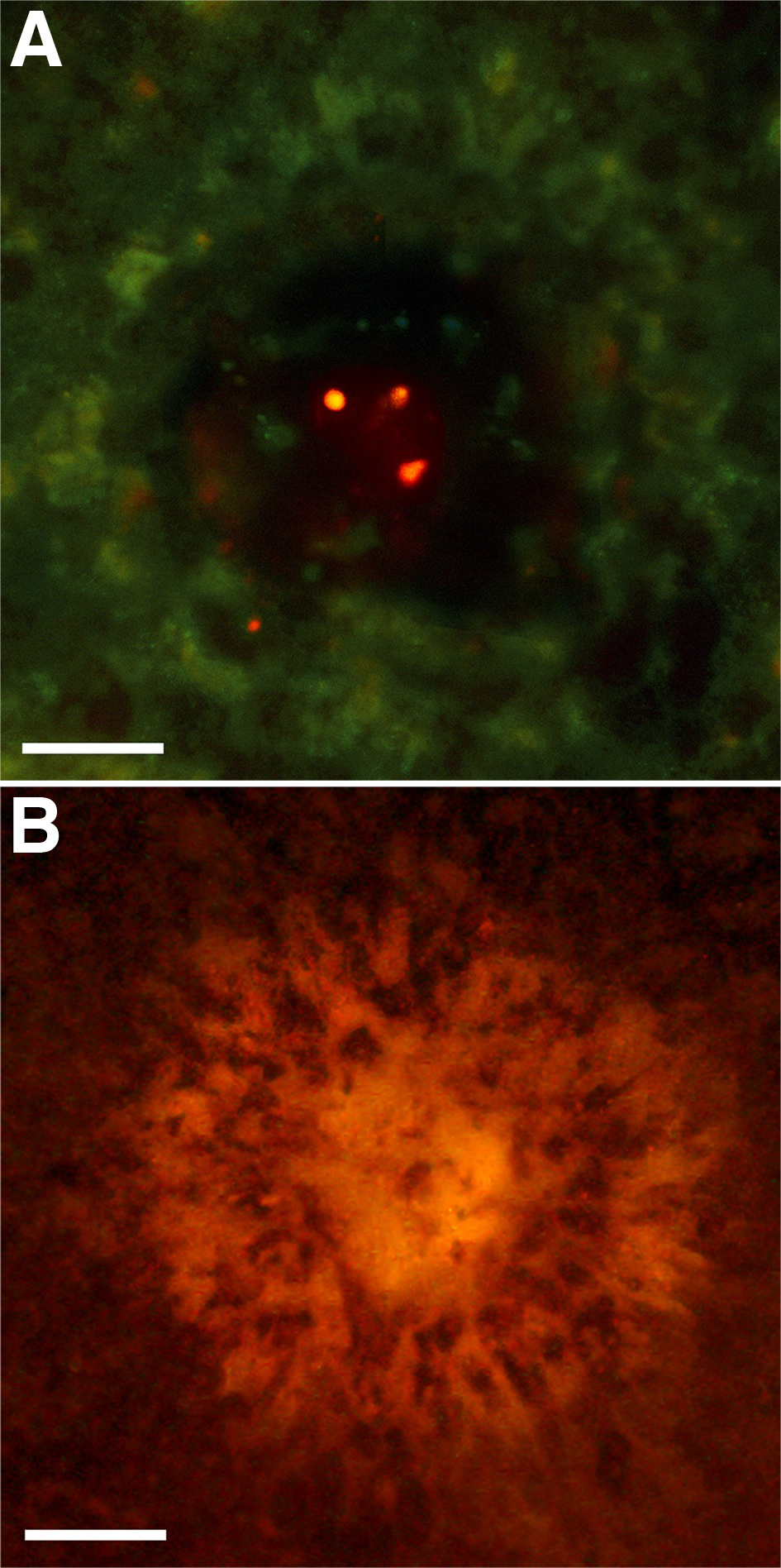
 Figure 2 of
Lundh von Leithner, Mol Vis 2010; 16:570-581.
Figure 2 of
Lundh von Leithner, Mol Vis 2010; 16:570-581.