Figure 1. In vivo and histological
fluorescence images exhibiting the impact of photocoagulation on
retinal pigment epithelium. A: In vivo near-infrared
reflectance image made immediately after photocoagulation (PC) showing
placement of central (arrowhead) and peripheral (arrow) lesions in the
retina. Scale bar represents 1 mm. B: In vivo autofluorescence
image of a lesion made 7 days after PC shows absence of fluorescence at
the center of the lesion. The lesion is surrounded by hyperfluorescent
point sources. Scale bar represents 200 µm. C: Autofluorescence
image of a lesion in retinal pigment epithelium (RPE), which was
excised and flatmounted 6 h after PC. The RPE tissue is continuous
across the lesion site, consistent with the notion that PC did not
result in immediate tissue destruction. D: Autofluorescence
image 72 h after PC, showing a clear lesion site and the absence of
tissue. Scale bars in C and D represent 50 µm.
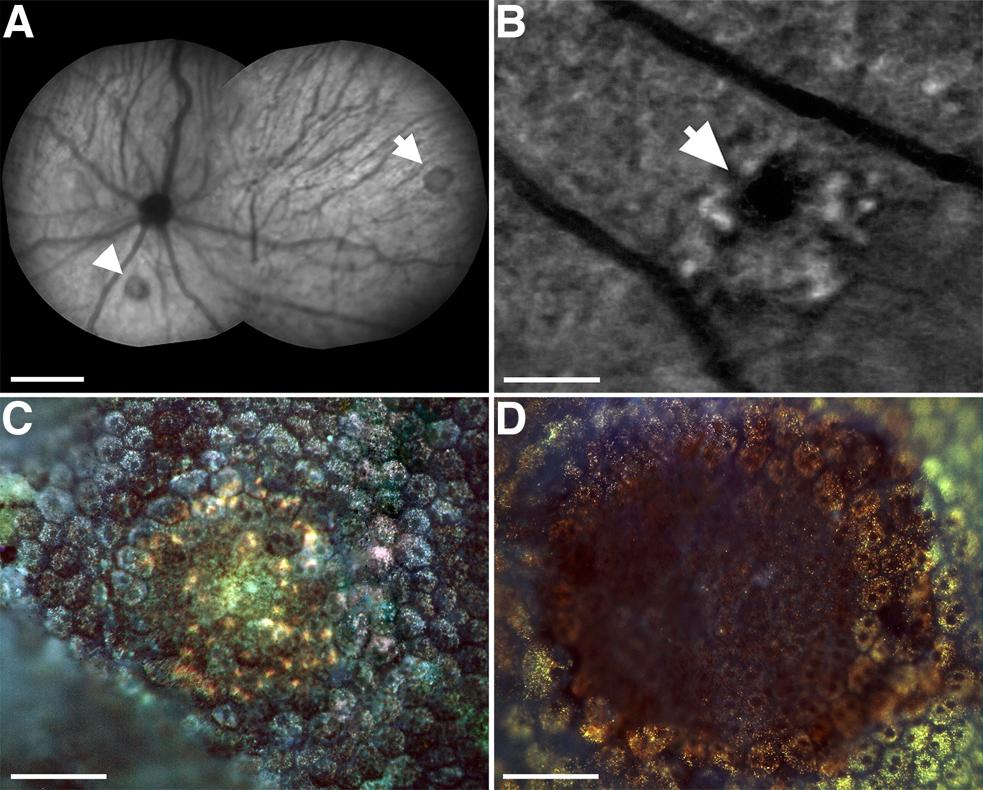
 Figure 1 of Lundh von Leithner, Mol Vis 2010; 16:570-581.