Figure 2. Slit-lamp examination showed an irregular corneal surface and several discrete, gray-white opacities in the subepithelial
area and Bowman’s layer. Some of the opacities were associated with marked corneal scarring. No neovascularization and stromal
lattice were observed.
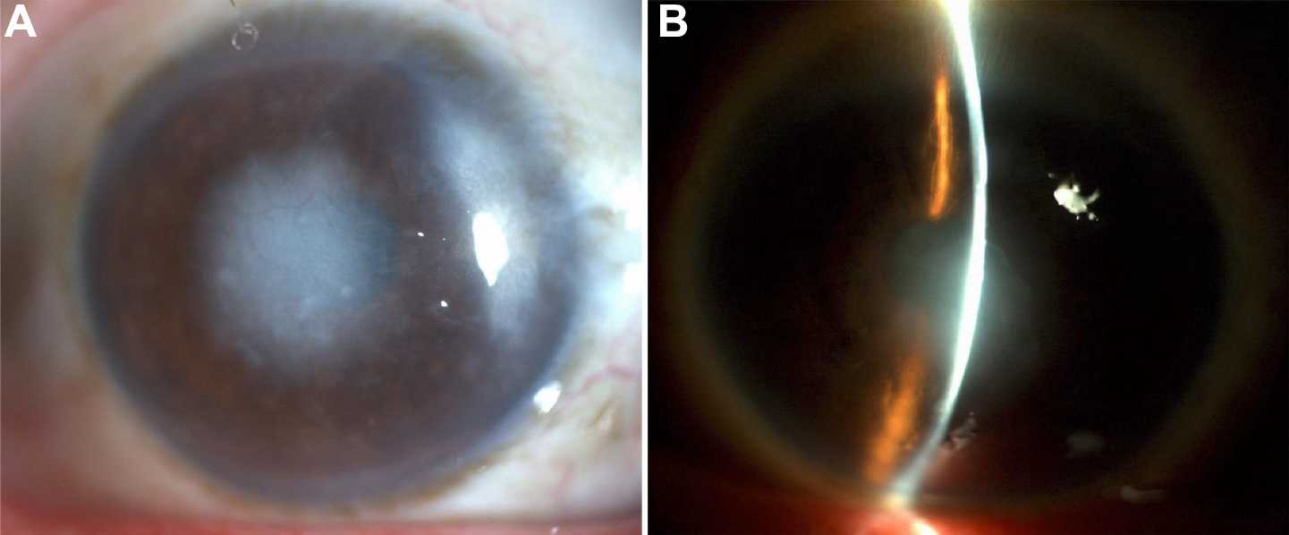
 Figure 2 of
Ma, Mol Vis 2010; 16:556-561.
Figure 2 of
Ma, Mol Vis 2010; 16:556-561.