Figure 2. Autofluorescence images and
high-resolution spectral domain optical coherence tomograms of affected
patients with membrane-type frizzled-related protein, and related
oculopathy. Autofluorescence images of the right (A) and left (B)
eyes
of posterior pole of individual III-1 of family 4 show relative
preservation of the central macula. Optical coherence tomograms of the
right (C) and left (D) fovea of individual II-1 of family
3 show areas of low reflectivity, which are due to cysts and relative
thickening of the retina in the foveal region.
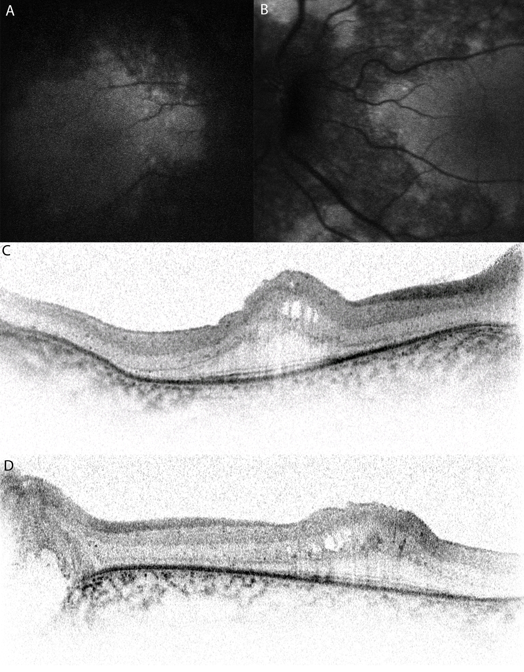
 Figure 2 of Mukhopadhyay, Mol Vis 2010; 16:540-548.