Figure 1. Color fundus photographs of
affected patients with membrane-type frizzled-related protein gene
related oculopathy. These show the right (A) and left (B)
fundus photographs of subject III-1 of family 4, the right fundus
photograph of individual II-1 of family 1 (C), and the left
fundus photograph of patient III-3 of family 2 (D); all show an
ellipsoid area of macula with normal reflex. Fundus photographs show
the right (E) and left (F) eye of individual III-3 of
family 2 after developing serous retinal detachments.
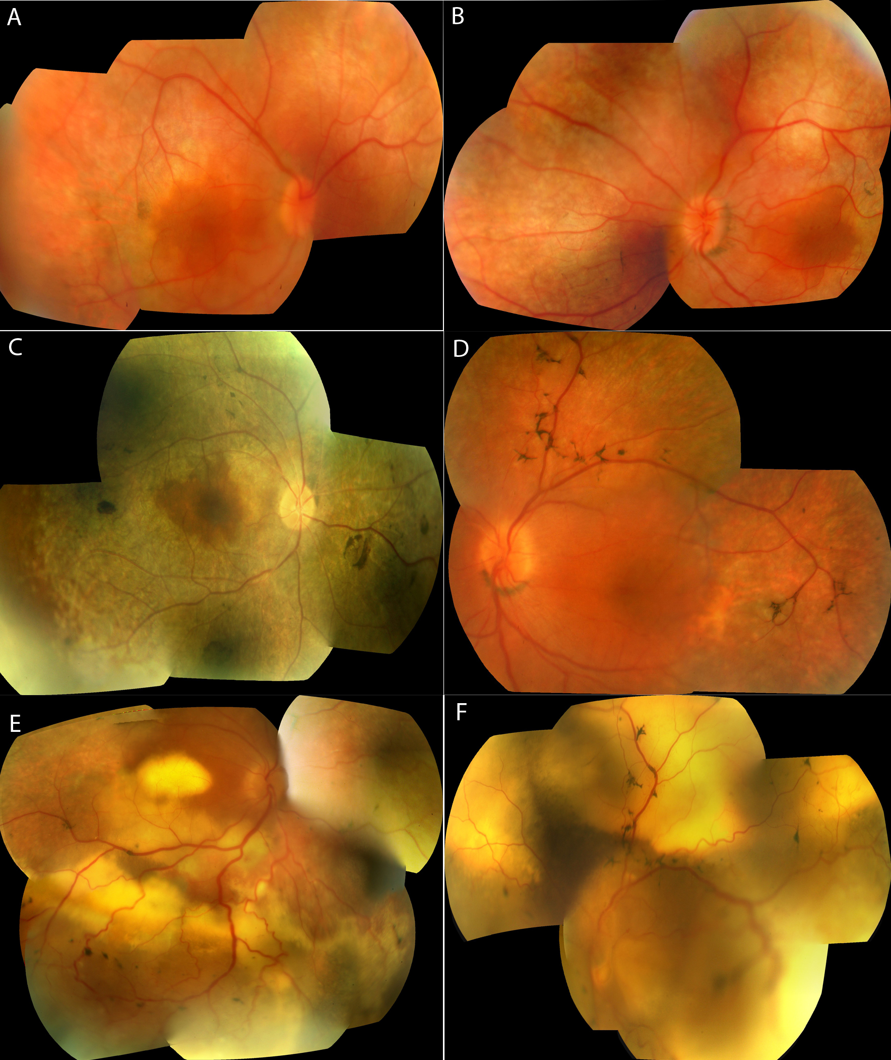
 Figure 1 of Mukhopadhyay, Mol Vis 2010; 16:540-548.