Figure 5. Color fundus composite
photographs of affected individuals from families 1 and 2 and Goldmann
visual fields from the proband in family 2. Panel A shows a
fundus composite of the right eye from 34-year-old male (IV:6) from
family 1; his visual acuity (VA) was 0.1 LogMAR with an
electroretinogram (ERG) that was consistent with a “mild cone–rod
dystrophy.” The image shows evidence of vessel attenuation and early
peripheral retinal atrophy with few bone spicules. Panel B
shows a fundus composite of the right eye in 36-year-old female (IV:5)
from family 1 whose VA was 0.3 LogMAR, and who had absent color vision;
her ERG was consistent with a diagnosis of retinitis pigmentosa. The
fundus image shows macular depigmentation, dense peripheral bone
spicules, and vessel attenuation. Panel C displays the color
fundus composite from the left eye of 42-year-old male (IV:3) from
family 1. His VA was 1.3 LogMAR, and the image displays circular
patches of RPE atrophy both at the macula and in the periphery with
associated peripheral pigment migration. His ERG was described as
consisted with “advanced RP.” Panel D shows a color fundus
composite from the right eye of 44-year-old female (IV:2) from family
1; Her visual acuity was limited to Counting Fingers (CF) and the view
of the posterior pole is obscured by dense asteroid hyalosis as seen;
there is peripheral bone spicule pigmentation and circular pigment
epithelial atrophy was observed in the far periphery. Panel E
shows the left eye color fundus composite of 46-year-old male (II:1)
from family 2. His VA was recorded as Hand Movements (HM) at his most
recent examination. No color vision was ever detected; the fundus image
shows an atrophic retina, with vessel attenuation, RPE atrophy around
the disc, at the macula and in the periphery, and associated peripheral
pigment migration. His ERG when tested at age 32 years was
undetectable. Panel F displays the Goldmann visual fields for
patient II:1 with the V4e isopter in green and III4e in red: there are
peripheral islands of residual vision and no response centrally to the
largest and brightest target (V4e) in the right eye and small areas of
response in the left eye.
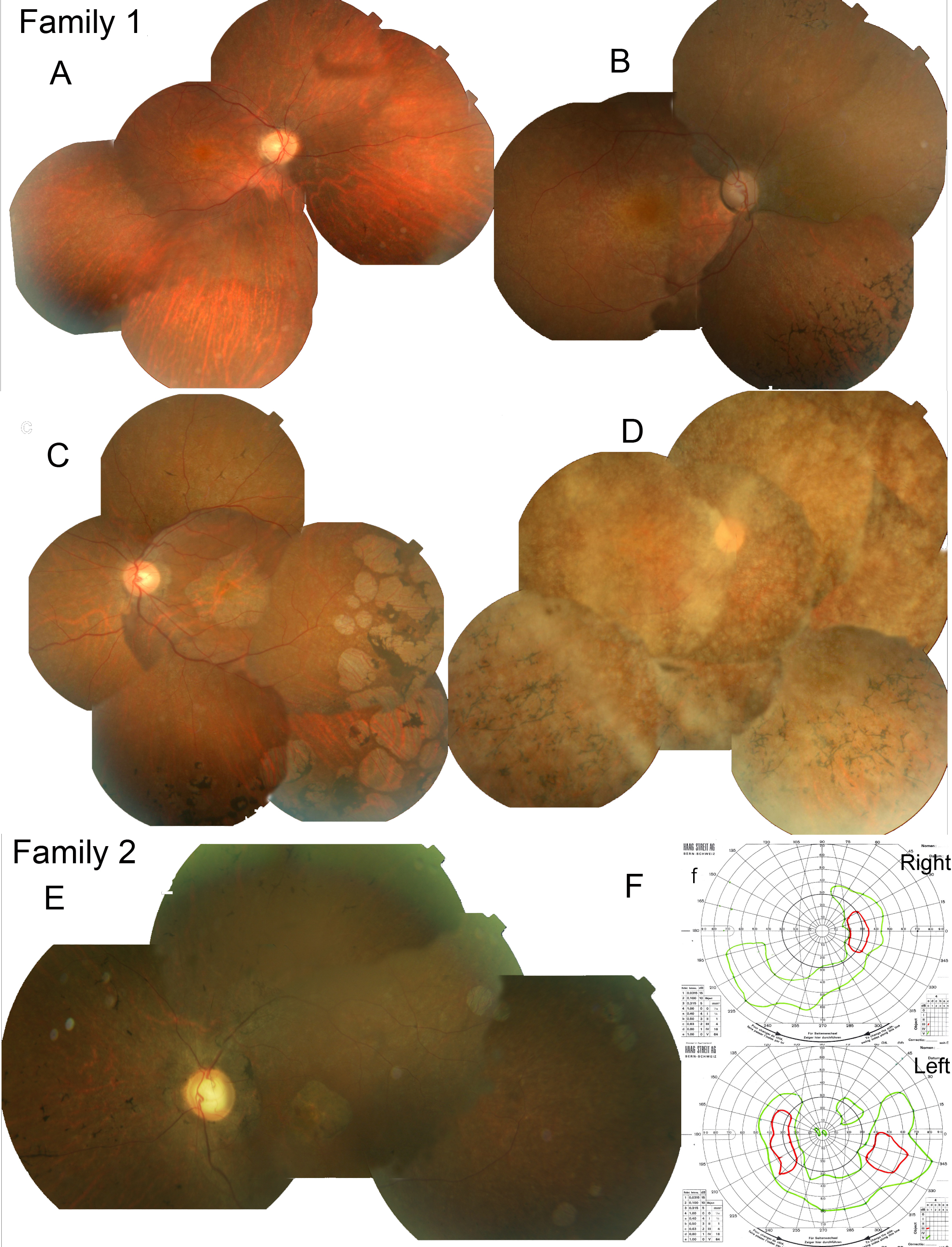
 Figure 5 of Henderson, Mol Vis 2010; 16:46-52.