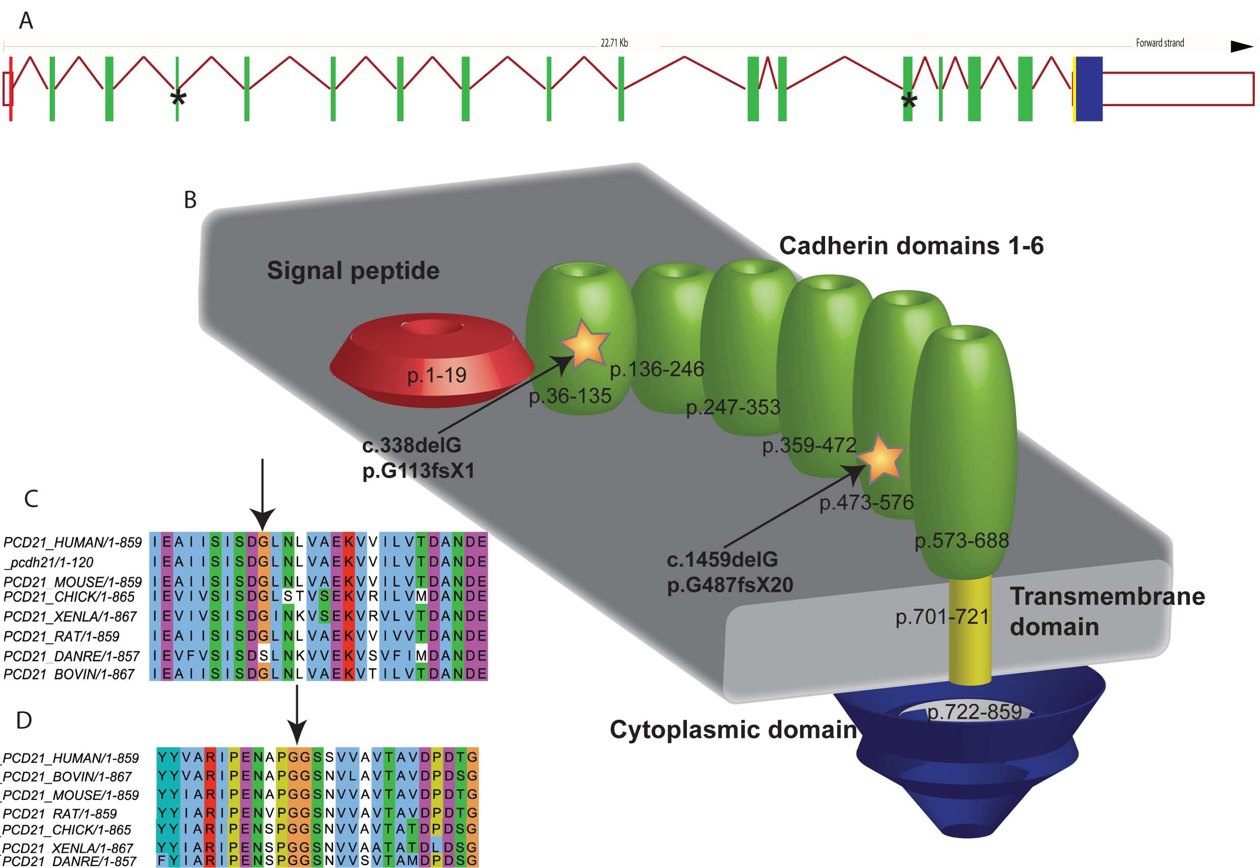
Figure 4. These are illustrations of both
the exon structure and a schematic of the Protocadherin-21 (PCDH21)
protein with indications of the location of the two deletions. Sequence
alignments demonstrate the conservation of both amino-acid positions
across several species. Panel
A: PCDH21 exon structure as
derived from
ensembl: the exons
are colored according to domain structure, and, as illustrated in Panel
B, with asterisks to denote the location of the novel deletions.
Panel
B is a schematic of the cellular domain structure derived
from
UniProt (created using Adobe
Illustrator-Adobe Systems Inc.) with the protein numbers corresponding
to each of the structural domains. Panel
C is an sequence
alignment created using PipeAlign for PCDH21 mutation p.G113fsX1, that
was identified in family 1, and shows that there is a high level of
conservation at this amino acid position-except for species
Danio
rerio. Panel
D is a further alignment around the site of
the PCDH21 deletion p.G487fsX20, identified in family 2, demonstrating
a high level of conservation at this amino acid position and therefore
providing evidence of the likely deleterious effect of this variant.
 Figure 4 of Henderson, Mol Vis 2010; 16:46-52.