Figure 2. Electropherograms of the
mutations detected in Protocadherin-21 (PCDH21). Panel A
illustrates the index c.338delG mutation in family 1; the wild-type
(WT) sequence is displayed on the top row. The middle row shows the
proband in family 1 (IV-2) with a homozygous (HM) c.338delG change,
illustrated with a dash for the missing nucleotide when aligned with
the wildtype sequence. The bottom row displays an unaffected parent of
family 1 (III-3) with the c.338delG change in the heterozygous state
(Het): the latter section of the heterozygous electropherogram shows
two superimposed sequences due to the synchronous addition of
nucleotides due to two distinct DNA templates derived from the wild
type and the shorter mutant alleles of the heterozygote. Panel B
illustrates the second mutation that was identified in PCDH21,
in family 2. The top row displays the control individual with the
wildtype (WT) allele; while the bottom row displays the affected
proband (II-1) with a homozygous (HM) c.1463delG variant.
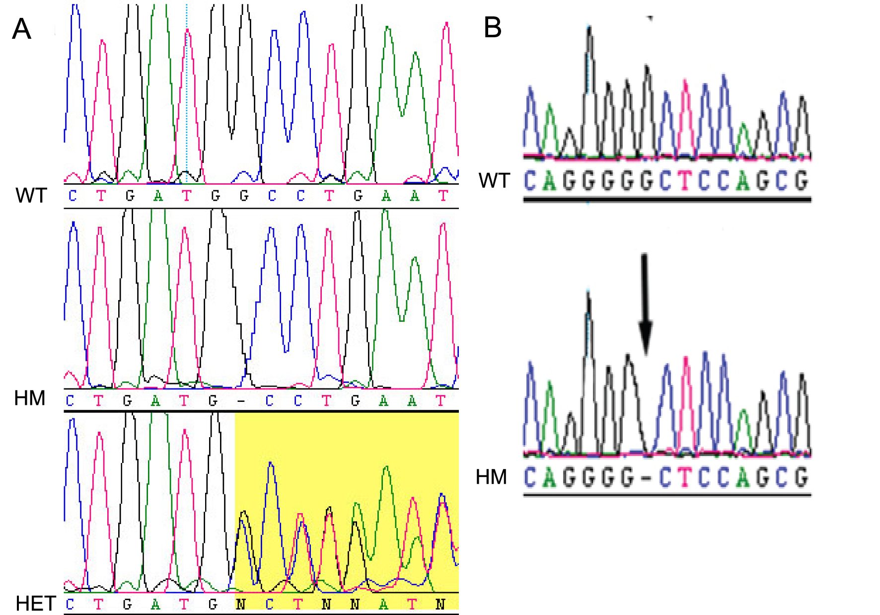
 Figure 2 of Henderson, Mol Vis 2010; 16:46-52.