Figure 5. Standard 2D gel of a 69-year-old
human lens. IEF was done in the first dimension using an 11 cm IPG
strip, pH 5–8, followed by SDS–PAGE using a 15% polyacrylamide gel in
the second dimension. The gel was stained with Coomassie blue R250, and
shows a profile similar to 2D-DIGE gels of the same tissue. However,
HMW (>35 kDa) aggregates are not distinguished as individual
spots, rather they appeared as non-descript bands.
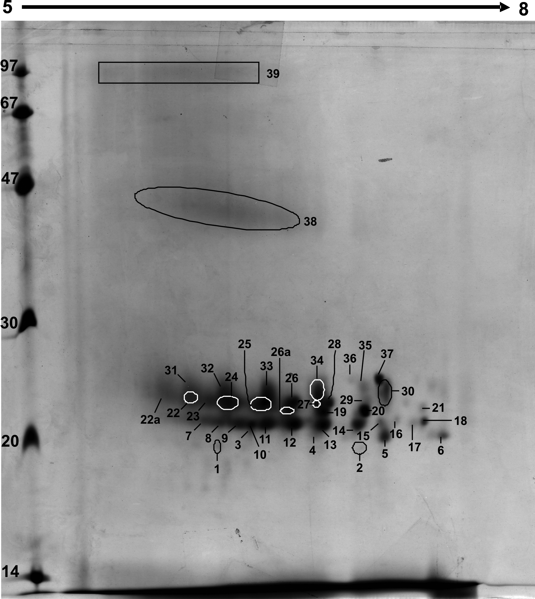
 Figure 5 of Asomugha, Mol Vis 2010; 16:476-494.