Figure 2. Separation of 65-year-old human
lens proteins using 15% polyacrylamide gel by SDS–PAGE analysis and
identification of excised bands by MALDI-TOF mass spectrometry. A:
SDS–PAGE
image of proteins in three lenticular regions with Coomassie
Blue R250 staining showing mostly LMW species. Samples from the outer
and inner cortices were repeated in the last two lanes on the right
side of the gel. B: Expanded image seen in A. Boxes
outline the bands excised for mass spectrometric analysis and
crystallin identifications of excised bands were based on MALDI-TOF
data.
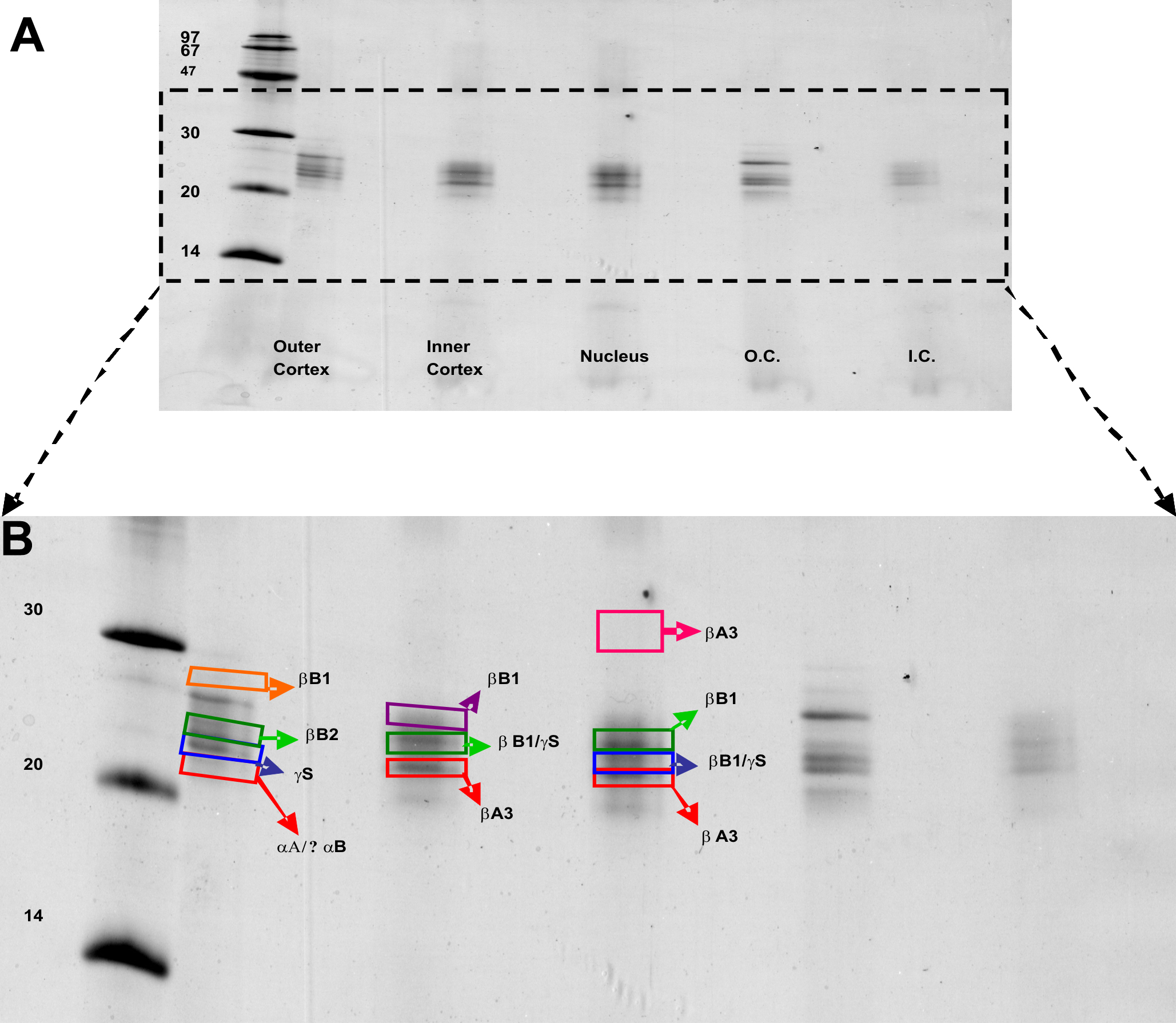
 Figure 2 of Asomugha, Mol Vis 2010; 16:476-494.