Figure 1. Tissue section of a human lens. A:
Whole
tissue section (12 μm) of 69-year-old lens stained with
hematoxylin and eosin, as seen under a stereomicroscope with 4×
magnification. All three major regions are present in this section,
however the nucleus stained more faintly because of possible increased
hydrophobicity of the proteins in this region. B: Unstained
section showing three major lenticular regions: Nucleus (N), Inner
Cortex (IC), and Outer Cortex (OC), as seen during LCM with the
Zeiss/PALM Microbeam microscope at its lowest magnification of 2.5×.
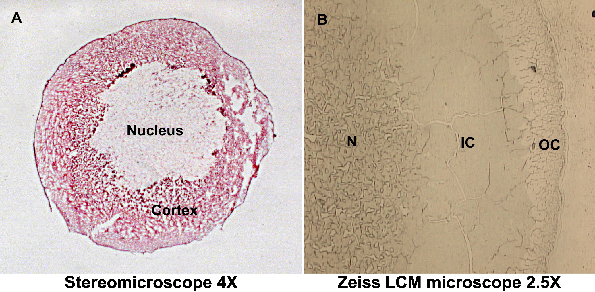
 Figure 1 of Asomugha, Mol Vis 2010; 16:476-494.