Figure 4. Autofluorescence and fundus photography. This image is showing a ring of high density autofluorescence imaging (AF) of a mother
A (IV.6) and her asymptomatic daughter C (V.10) with normal fundus. A: An AF image of 39-year-old mother, left eye. Macula with a hyperfluorescent ring. (B) Color photo of the same patient. Slight narrowing of retinal arteries. C: An AF image of her 10-year-old asymptomatic daughter, left eye. Macula with a large hyperfluorescent ring D: Color photo of the same patient. Normal fundus.
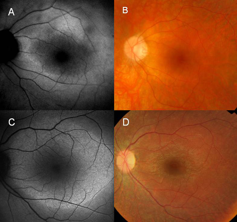
 Figure 4 of
Vaclavik, Mol Vis 2010; 16:467-475.
Figure 4 of
Vaclavik, Mol Vis 2010; 16:467-475.