Figure 3. Autofluorescence and fundus photography. This image is showing a ring of high density autofluorescence of a daughter, A (V.3) with normal fundus and her father C (IV.3). A: An autofluorescence imaging (AF) image of four-year-old girl, right eye. Macula with a large hyperfluorescent ring. B: Color photo of the same patient. Pale appearance of fundus is shown. C: An AF image of her 44-year-old father, left eye. Macula with a small hyperfluorescent ring. D: Color photo of the same patient. Pale disc, narrowed retinal vessels, bone spikes at close periphery are shown.
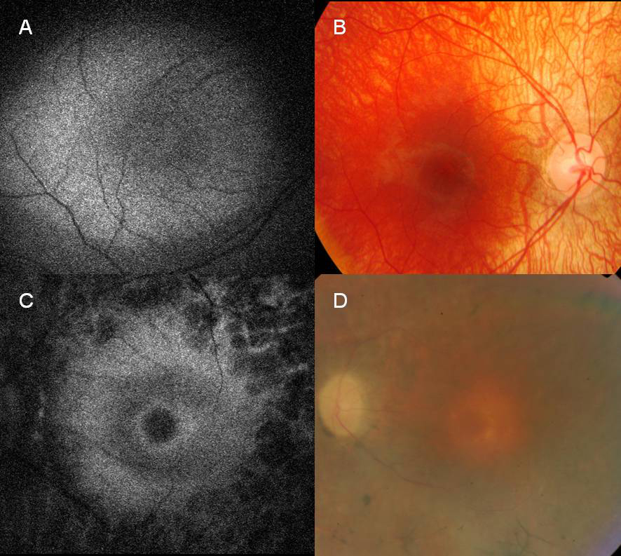
 Figure 3 of
Vaclavik, Mol Vis 2010; 16:467-475.
Figure 3 of
Vaclavik, Mol Vis 2010; 16:467-475.