Figure 2. Autofluorescence and fundus photography. This image is showing progressive constriction of perifoveal ring of high density
autofluorescence in the grand-mother A (Proband III.9), her daughter C (IV.9) and the grand son D (V.11). A: An autofluorescence imaging (AF) image of 70-year-old mother, right eye. Macula with a small hyperfluorescent ring. B: Color photo of the same patient. Pale disc, bones spikes at the periphery are shown. C: An AF image of the right eye of her daughter, 46-year-old. Macula with a larger hyperfluorescent ring. D: Color photo of the same patient. Normal appearance is shown. E: An AF image right eye (RE) of 22-year-old grandson. Macula with a larger hyperfluorescent ring. F: Color photo of the same patient. Normal appearance is shown.
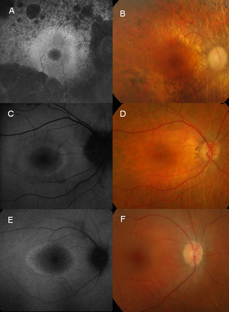
 Figure 2 of
Vaclavik, Mol Vis 2010; 16:467-475.
Figure 2 of
Vaclavik, Mol Vis 2010; 16:467-475.