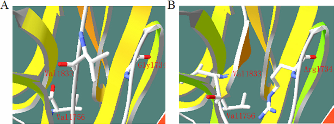
Figure 5. 3D modeling of p.G1734R (PDB template 2JD4_B, 26% identity with Lama1).
A: wild-type protein,
B: mutant protein. The replacement of Gly by Arg may induce a new secondary structure that includes Leu1831, Val1832, and Val1833.
The new secondary structure makes Val1833 very close to Val1756, which may change the conformation of the binding site. The
structure of the wild type and the mutant USH2A protein were predicted using
Swiss Pdb-Viewer 4.0.1.
 Figure 5 of
Liu, Mol Vis 2010; 16:454-461.
Figure 5 of
Liu, Mol Vis 2010; 16:454-461.