Figure 4. Identification of two novel USH2A
mutations. DNA sequence analysis for patient III1 showed the presence
of compound heterozygous p.G1734R (c.5200G>C) and c.IVS32+1A>T
mutations. A and B show the sequences of a normal and
affected family member with mutation p.G1734R (c.5200G>C) allele,
respectively. C and D show the sequences of a normal
and affected family member with mutation c.IVS32+1A>T allele,
respectively. E: Restriction fragment length analysis on the
p.G1734R (c.5200G>C) mutation in this study. All the affected
individuals (III:1, III:3, III:5, IV:1, IV:2) and the carriers (III:2,
IV:3) have three bands (228 bp, 164 bp, and 64 bp), while the
unaffected individuals only have two bands (164 bp and 64 bp).The
patient IV1 who is homozygous for the p.G1734R (c.5200G>C) mutation
displayed only one 228 bp band. F: Alignment of the amino acid
sequences of laminin G-like domain in the long usherin isoform from
different species. Gly1734 (G1734) is conserved during evolution. The
box indicates this mutated residue in USH2A.
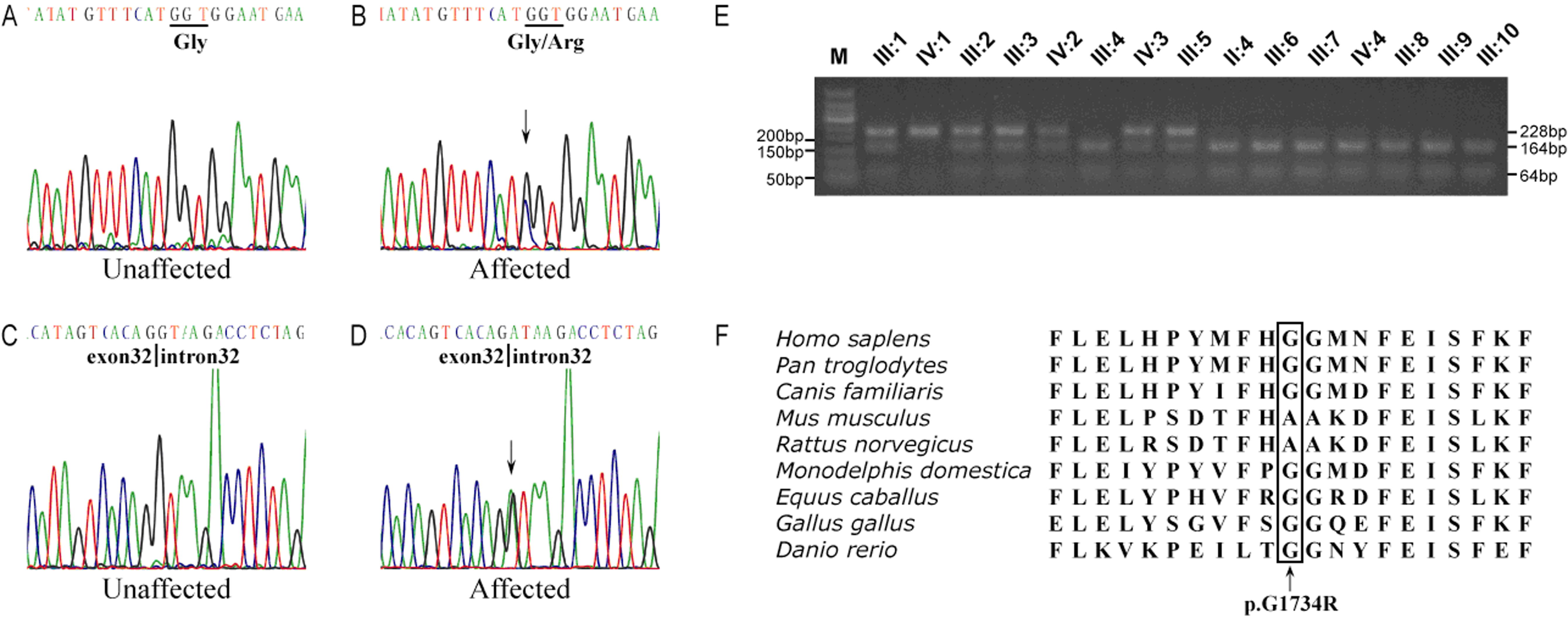
 Figure 4 of Liu, Mol Vis 2010; 16:454-461.